


Search results for 'lang'
-

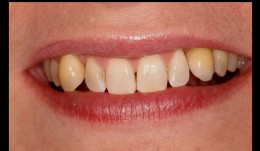
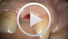
Periodontitis case, single implant crown
Prof. Niklaus Lang and J. TamMale patient *1970, by J. Tam and N. P. Lang (2011-2013), University of Hongkong. Periodontitis case with pain and furcation involvement, single implant crown -
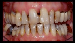
Periodontitis case, smoker, shortened dental arch, reconstructions on implants
Prof N. P. Lang and M. Lulic.Female patient *1951 by M. Lulic. N. P. Lang (2006-2008). Periodontitis case, smoker, shortened dental arch, reconstructions on implants (3-unit bridge and single crown). -
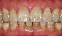
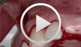
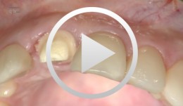
Periodontitis case, smoker, single anterior implant crown
Prof N. P. Lang and M. Lulic.Female patient *1966, by M. Lulic and N. P. Lang (2007-2008). Periodontitis case with pain, smoker, single anterior implant crown. -
Regenerative Behandlung multipler Rezessionen
Heinz, BerndGliederung - Befund - Schnittführung - Wurzelglättung - Applikation von PrefGel - Applikation von Emdogain - Periostschlitzung - Nahttechnik Inhalt: Regenerative Parodontalchirurgie mit dem Schmelz-Matrix-Protein Emdogain: Ziel der regenerativen Parodontalchirurgie ist der Wiederaufbau zerstörter parodontaler Strukturen. Eingesetzt wurden bislang und werden nach wie vor Knochentransplantate, Knochenersatzmaterialien sowie nicht resorbierbare und resorbierbare Membranen. Ein neuerer therapeutischer Ansatz zur parodontalen Regeneration ist die Wurzeloberflächenkonditionierung mit dem Schmelz-Matrix-Protein Emdogain (Biora, Schweden). Durch den Proteinkomplex wird die Neubildung von Wurzelzement stimuliert, was wiederum die Neubildung von Knochen und Kollagenfasern zur Folge hat. Zu diesem Verfahren und seiner Wirkungsweise wurden seit Anfang 1980 umfangreiche Untersuchungen und Studien von einem schwedischen Forscherteam um Prof. Lars Hammerström durchgeführt. Mittlerweile wird das Schmelz-Matrix-Protein Emdogain zur Behandlung von vertikalen Knochendefekten und von Furkationserkrankungen eingesetzt. Die freigelegte Wurzeloberfläche wird zunächst sorgfältig mit Handinstrumenten oder rotierenden feinkörnigen Diamanten gescalet. Sodann wird die EDTA-Suspension Pref-Gel (Biora, Schweden) aufgetragen und nach zwei Minuten mit physiologischer Kochsalzlösung gründlich abgespült. Die EDTA-Suspension bewirkt die Entfernung des Smearlayers und öffnet die Dentintubuli, wodurch eine bessere Verbindung von Emdogain an die Wurzeloberfläche erreicht wird. Sofort anschließend wird Emdogain auf die blut- und speichelfreie Wurzeloberfläche appliziert. Danach erfolgt der Nahtverschluss. -
Insertion von zwei enossalen Implantaten Regio 11-21
Hildebrand, DetlefGliederung: - Schnittführung - Lappenpräparation - Applikation der Bohrschablone - Bohrung - Implantation - Applikation der Abdruckpfosten - Naht - Einsetzen des Provisoriums Materialliste: Camlog RootLine Implantate, Ø 4,3mm, 13 mm Länge; E-Woo Picasso Trio: Digitales 3-D Imaging System (3in1) -
Sofortimplantation Regio 12 und 22
Diemer, JosefGliederung - Schnittführung regio 22 - Lappenpräparation - Bohrung - Implantatbettaufbereitung - Implantation - Applikation der Verschlussschraube - Naht - Extraktion Wurzelrest regio 12 - Kürretage - Implantation - Applikation der Verschlussschraube Materialliste Implantate: 3I, Osseotite, Aussenhex, Durchmesser 4mm. Rg. 12 mit Länge 15mm Rg. 22 mit Länge 13mm -
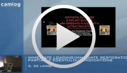
Immediate loading/immediate restoration - partially edentulous indications
de Lange, GertHighlights of the International CAMLOG Congress 2008 9. und 10. Mai 2008 in Basel, Schweiz www.camlog.com -
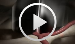
Kommunikation der Zellen - Die Osseointegration (Trailer)
Stadlinger, Bernd / Terheyden, HendrikDas Unsichtbare sichtbar werden zu lassen - darin liegen die Faszination und die Herausforderung, die heute bekannten zellbiologischen Hintergründe der Osseointegration anhand der beteiligten Zelltypen und Botenstoffe zu visualisieren und diese komplexen biodynamischen Prozesse dramaturgisch und didaktisch so zu gestalten, dass sie in der Aus-, Fort- und Weiterbildung eine wertvolle Unterstützung in der Wissensvermittlung bieten. Mit dem Modul 1 "Kommunikation der Zellen - Die Osseointegration" startet die Exzellenzinitiative "Lehre - Lebendige Wissenschaft", in der sukzessiv alle relevanten biomedizinischen Prozesse in der ZMK als 3D-Computerfilmanimationen produziert und in einer 3D-Filmbibliothek der zahnmedizinischen Fachwelt zur Verfügung gestellt werden. Dieses neue Genre bietet interessante Perspektiven für die Lehre und ein Highlight für den Betrachter. Gliederung: - Die Hämostase - Die entzündliche Phase - Die proliferative Phase - Die Remodellierungsphase Zum Film Hauptdarsteller: Thrombozyten, Fibroblasten, Endothelzellen, Granulozyten, Makrophagen, Perizyten, Osteoklasten, Osteoblasten, Osteozyten Nebendarsteller: PDGF, Thromboxan, TGF-a, TGF-ß, PDGF, VEGF, NO, ACE, TNF-a, IL-1, TNF-a, IL-6, FGF, MIP-1, RANKL, Sclerostin Filmlänge: zwölf Minuten Das Projekt- und Expertenteam Wissenschaftliche Leitung: Dr. Dr. Bernd Stadlinger, Prof. Dr. Dr. Hendrik Terheyden Advisory Board: Prof. Dr. Christoph Hämmerle, Prof. Dr. Thomas Hoffmann Fachliche Beratung: Dr. Susanne Bierbaum, Prof. Dr. Dr. Uwe Eckelt, Dr. Ute Hempel, Prof. Dr. Lorenz Hofbauer, Prof. Dr. Dieter Scharnweber (Transregio 67) -
Sofortimplantation und Versorgung 21 in Kombination mit autologem Knochenaufbau der bukkalen Wand
Körner, GerdGliederung: - Zahnentfernung 21 - Säuberung und Darstellung der defizitären Alveole - Präparation und Implantat Kavität (Beginn mit Facilitate®, individuelle Nachbearbeitung) - Implantateinbringung - Gewinnung des autologen Knochens aus Linea obliqua - Einbringen des partikulierten Knochens in das bukkale Knochendefizit - Anfertigen der prothetischen Sofortversorgung - Eingliedern der adhäsiv befestigten Sofortversorgung. Materialliste: - Facilitate® Bohrschablone (entsprechend der DVT Auswertung) - Astra Profile Implantat ø 4,5 mm, Länge 15 mm - Astra Zebra Abutment - Astra Ti - Unite Profile - Astra Lab - Analog ø 4,5 mm - Voco Struktur - Relay - X - Veneer - Temp - Bond (Kera) -

ANTIBODY-MEDIATED OSSEOUS REGENERATION (AMOR) IN THE RECONSTRUCTION OF ALVEOLAR RIDGE DEFECTS
Objectives: The aim of this study was to examine the efficacy of antibody-mediated osseous regeneration (AMOR) in conjunction with two novel extraction devices, namely a SocketKAP™ (for obturation of socket orifices) and SocketKAGE™ (for providing space in sites with facial dehiscence for ridge preservation and augmentation procedures following tooth extraction). Methods: In this prospective controlled study, 8–12-year-old macaques (Macaca fascicularis; n = 6) underwent removal of both maxillary and mandibular second left and right premolars (ULPM2, URPM2, LLPM2, LRPM2), both maxillary and mandibular second left and right molars (ULM2, URM2, LLM2, and LRM2) on each side. This was followed by resection of the entire facial alveolar wall. They were randomly assigned to the following intervention groups: dehiscence socket unfilled and uncovered (group A; negative control); dehiscence socket reconstructed with SocketKAGE™ and covered with SocketKAP™ (group B); dehiscence socket reconstructed with SocketKAGE™ and ABBM and covered with SocketKAP™(group C); dehiscence socket reconstructed with SocketKAGE™ and chimeric anti-BMP-2 monoclonal antibody (mAb) and covered with SocketKAP™(group D); dehiscence socket reconstructed with SocketKAGE™ and iso-type-matched control mAb (Iso-mAb) and covered with SocketKAP™ (group E). At 6 and 12 weeks post-extraction, cone beam CT imaging was performed. All files were imported to Simplant TM/® software (for linear measurement of remaining bone width and height at 1mm, 2mm, 3mm and 5mm from alveolar crest) and to reverse engineering Geomagic ControlTM/®software (for volumetric analysis of alveolar bone). Histologic evaluation was also made of the dimensional changes in dehiscence defects after ridge preservation using different materials. Non-parametric analysis of variance was conducted using the methods of Brunnner and Langer. Results: The result from linear and volumetric analyses demonstrated that group D (chimeric mAb +SocketKAP™ + SocketKAGE™) was associated with significantly greater percentages of remaining bone width and height at the buccal point, and remaining bone volume at 6 and 12 weeks when compared to group A (negative control), group B (SocketKAP™ + SocketKAGE™) and group E (Iso-mAb); the latter lost approximately 70% of crestal bone width at the crestal 1 mm point, and ABBM failed to prove more effective in preserving alveolar bone width at crestal 1mm (36%). When anti-BMP-2 mAB was used, the percentage of bone width was as good as those with ABBM at crestal 2mm, 3mm and 5mm areas. However, the most important finding was that the data demonstrated that bone remodelling using chimeric mAb was not only limited to the crestal 2–5-mm zone, but also the crestal 1-mm area, with mean remaining bone widths of 52% and 68% at 6 weeks and 12 weeks, respectively. Both CB-CT and volumetric analysis revealed more active and extensive bone regeneration of marginal bone with chimeric mAb compared to other groups. Histologic analysis showed the knife-edge of alveolar crest following tooth extraction in groups A, B, C and E, as well as statistically significant more bone remodelling in chimeric mAb, including a higher percentage of bone formation within newly generated tissue together with signs of osteogenic cell activity. Conclusions: These results suggest that chimeric anti-BMP-2 mAb, in conjunction with SocketKAPTM/® and SocketKAGETM/® was effective in the reconstruction of the alveolar bone in extraction sockets with facial dehiscence defects. These favourable results may be attributed to advantageous nature of AMOR, such as using endogenous BMP-2 captured by chimeric mAb enhance osteogenesis, with higher biological activity of endogenous BMP and a longer half-life of captured BMP compared to ABBM (which only exhibits osteoconductivity).