


-
- English
- Deutsch
- Spanish
-
- General Diagnostics
- Treatment
- Tools/Materials
- Basic/General Knowledge
- Research
- Events
- Organisations
-
-
-
-
-
-

-


Short and narrow implants, how far can we go?
Christoph Hämmerle, José NartIn this webinar moderated by Prof Ronald Jung and Dr. Adrián Guerrero the expert presenters Prof. Christoph Hämmerle and Dr. José Nart discuss about the importance and benefits of using short and narrow implants. -

-

THE OSTEOLOGY CASE BOX
The Osteology CASE BOX contains cases uploaded for different clinical scenarios. User decides whether they want to share their cases, or whether they only add the data for overall evaluation and compare it with the mean. -
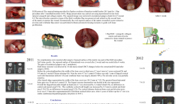
ALVEOLAR RIDGE PRESERVATION IN DAMAGED SITE 4.7—AN EARLY STAGED OPEN-HEALING PROTOCOL IN HIGH-RISK PATIENTS TREATED WITH ORAL BISPHOSPHONATES
Objectives: The objective was to simplify the regenerative procedure using a minimally invasive technique. Ridge preservation at the time of the patient’s tooth avulsion was not possible because of acute infection. Surgery was performed using biomaterials during the early post-extractive healing process. The "early build-up technique", as used in healthy patients, was chosen to reduce comorbidity risks in a single high-risk patient. Methods: A 69-year-old woman had been treated with oral alendronate 70 mg weekly since 2001 because of a vertebral fracture caused by osteoporosis. She had experienced in anamnesis breast neoplasm in 2000 (and recurrence in 2015) and myocardial infarction for which she had a triple bypass in 2007 and angioplasty in 2014. The patient attended the clinic for chronic periodontitis and the tooth 4.7 was extracted because of recurrent periodontal abscesses. Before the extraction, cone beam computed tomography (CBCT) allowed evaluation of cortical wall thickness around the mobile mesial inclined 4.7 from the most significant eight cross sections (CS), and 16-mm mesiodistal bone assessment. The edentulous area 4.6 was partially included in the evaluation. Photographs were taken and periodontal probing performed 2 months after extraction of 4.7. The surgical protocol involved flapless avulsion of the tooth in October 2011 and the flap procedure 2 months later (December 2011). The early alveolar build-up was performed using bovine-derived xenograft and collagen matrix. The surgical design was restricted to keratinised gingiva 4.6–4.7. The intra-alveolar connective tissue of the thick vestibular flap was preserved and rotated on the second layer of the matrix to protect the wound. The exposed surface of the matrix resembled geometrically a post-extract alveolus and flap suture was performed without periosteal-releasing incisions to guide soft tissue proliferation (GGP; guided gingival proliferation). Results: No complications were reported after surgery. The exposed matrix surface was part of the GGP procedure. As in healthy patients, the exposed biomaterial surface was sealed after 4 weeks. The gain in keratinised gingiva was 2mm. The hard tissue outcome was detected by 16-mm mesiodistal CBCT changes before the extraction and after surgery. Before the extraction, the bone crest width in the distal area 4.7 was 6 mm in two cross-sections and 8–9 mm in three mesial cross sections (area missing 4.6). Near the roots in 4.7 in three central cross-sections, there was only 1 mm of lingual cortical wall with fenestration defects (1 CS) and vestibular bone was largely absent (>50%). The alveolar cavity was partially retentive. After lateral ridge regeneration the bone crest width was 10–12 mm in seven cross-sections, with the greatest linear horizontal bone gain (of 10 mm) in four central cross-sections. The lingual osseous fenestration was healed. Overall the lingual bone plate height in distal area 4.7 was decreased by 1–2 mm (three distal cross-sections), with no differences in central and mesial areas (five sections). The vestibular cortical wall height was 2–5 mm greater in the central and distal area 4.7 (six cross-sections), with no differences in the mesial area (two cross-sections). The vertical distance between bone crest and alveolar nerve level was 8–9 mm in all cross sections, without residual defect area. After 4 years of follow-up the 4.7 site is in a stable condition according to ortopantomography and photographs. Conclusions: In healthy patients, this surgical augmentation of horizontal alveolar bone and keratinised gingiva is problem-free, and was shown to be the case also in this high-risk patient, in whom implant surgery is contraindicated because of comorbid conditions and uncontrolled periodontitis. In healthy patients, implant insertion is possible 6–12 months after bone augmentation. The choice of the GGP protocol for this alendronate-treated patient was associated with fewer risks than surgery with primary intention healing of the wound. Further clinical trials should be conducted on the GGP protocol, and may show that covering bone substitutes with collagen matrix exposed during the early post-extractive wound healing process is a surgical step instead of a complication of the GBR procedure. -

REAL-TIME NAVIGATION: THE BEGINNING OF A NEW ERA IN GUIDED IMPLANT SURGERY
Objectives: To demonstrate that dynamic guided surgery is as predictable as conventional surgery. Methods: Partially edentulous patients requiring a fixed rehabilitation were selected for this pilot study. No specific contraindications were established, and smokers were not excluded. An impression was taken pre-operatively using an irreversible hydrocolloid (Cavex CA37®) to fabricate a diagnostic cast for moulding the surgical stent (NaviStent®). Afterwards, a standard cone-beam CT (CBCT) scan was made with the NaviStent® in place using a Planmeca Promax 3-D Max®. Images were converted into DICOM files and transformed into a 3-D virtual model using the Navident® software. The potential implant locations were planned in a prosthesis-driven way. For preparing the osteotomy, the drilling axis of the handpiece and the twist drills were calibrated. The osteotomies were prepared at low speed using a high level of cooling. The navigation software guided the drilling procedure in real time. Before installing implants, an extra calibration procedure was performed for tracking the implant. The aim of this pilot study was to determine the clinical outcome up to 12 months post-operatively for implants installed using the Navident® guided surgery system. Results: Partially edentulous men (n = 6) and women (n = 7) were included in this pilot study (mean age 52.15 years; range 20–75). Out of these 13 patients, two were current smokers of more than 10 cigarettes per day. Twenty implants were inserted. No mechanical or biological complications occurred during the surgical procedure, and no major complaints were reported, such as hemorrhage, sinus pathology or severe post-operative pain. No implants were lost up to 1 year after insertion, resulting in 100% implant survival. Conclusions: Based on the results of this pilot study, real-time navigation is a promising technique. However, there is not yet enough evidence to show that the method is as safe and predictable as conventional implant surgery. -

HYBRID SCAFFOLDS COMPOSED OF BETA-TRICALCIUM PHOSPHATE (TCP), poly (D,L- lactic acid) (PDLLA) AND COLLAGEN FOR ALVEOLAR BONE AUGMENTATION
Objectives: Dimensional changes occur in the alveolar ridge after tooth loss, due to bone resorption. Bioactive ceramic scaffolds are available for bone alveolar augmentation, but their mechanical properties are still an issue during implantation. The aim of this study was to characterize the mechanical properties of synthesized β-TCP hybrid scaffolds coated with bioabsorbable collagen or PDLLA. Methods: β-TCP powder was obtained by reactive milling. Scaffolds were produced by the replica method from polyurethane foam patterns. Type I collagen or poly (D,L- lactic acid) (PDLLA) were used to coat the scaffolds by dip coating. The hybrid scaffolds were then divided into four groups: non-coated (GA); double immersion in collagen type I (GB); double immersion in PDLLA (GC); and ten immersions in PDLLA (GD). Samples were characterized by compressive tests, x-ray diffraction and scanning electron microscopy with energy dispersive x-ray analysis (SEM/EDS). Statistical analysis was performed by two-way ANOVA(p Results: Chemical and microscopic analyses revealed proper morphology of powder particles and scaffolds with or without polymeric coatings. Hybrid scaffolds of PDLLA had higher compressive strength (0.11 MPa ± 0.054) (p Conclusion: β-TCP scaffolds synthesised by the replica method showed proper morphology and size of interconnected pores. The PDLLA scaffold-coating method increased mechanical strength of the porous ceramic material, showing desirable properties such as high compressive strength, biocompatibility and osteoconductivity. These properties are essential in alveolar preservation procedures for resisting compression from soft tissues and occlusal stress from mastication. -
STEM CELL BONE ALLOGRAFTS IN MAXILLARY SINUS AND RIDGE AUGMENTATION – REPORT OF A CASE
Objectives: To evaluate the use of an allograft cellular matrix containing live stem cells for maxillary sinus and ridge augmentations. Methods: Maxillary sinus and ridge augmentations were performed using an allograft cellular matrix containing live stem cells. The post-operative results were evaluated by CT scans and peri-apical radiographs. Sinus augmentation was evaluated after 10 weeks. Radiographic bone tomography was similar to that of the native bone and the ridge augmentation resulted in a vertical ridge augmentation of 3–4mm. The cellular matrix was supplied by Brockton, MA and processed by AlloSource, Centennial, CO. Results: Following healing and approximately 10 weeks following surgery, an additional CT scan was taken. This showed that the native and augmented bone was of an adequate width for supporting an implant. Radiography revealed that the augmented bone had a similar texture to native bone, indicating formation of mature bone. The scan also revealed downward growth of the bone in a vertical direction, overlapping the crest of the native pre-maxillary bone. This was not attempted during the surgical procedure, and was a particular cause for concern. Conclusions: This use of allograft mesenchymal stem cells has been shown to be a reliable method for ridge augmentation, especially in the vertical direction in areas of severe ridge atrophy. Further studies are needed to support this finding in a more guided manner, especially for vertical ridge augmentation. -
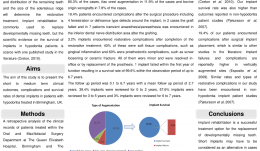
DENTAL IMPLANT SURVIVAL IN HYPODONTIA PATIENTS: A RETROSPECTIVE STUDY
Objectives: Specialists in prosthodontics and restorative dentistry are often challenged by oral rehabilitation of patients with hypodontia. Implant rehabilitation is commonly used to replace developmentally missing teeth, but evidence of implant survival in hypodontia patients is scarce. The aim of this study was to present findings on short- to medium-term clinical outcomes, complications and survival rates of dental implants in patients with hypodontia who were treated in Birmingham, UK. Methods: A retrospective analysis was conducted of the clinical records of patients treated within the Oral and Maxillofacial Surgery Department at Queen Elizabeth Hospital and the Restorative Dentistry Department at the University of Birmingham Dental Hospital. All patients were identified from the hypodontia clinic database. Clinical variables were recorded, collated and compared with reports of adverse events during a follow-up period of up to 6.7 years. Patient data were collected using a pro forma regarding the number and the location of implants, the surgical procedure, and any surgical or restorative complications. All patients on the study had a review appointment within the previous 3 months. The data were analysed to determine the number of patients treated, the type and number of implants placed, the site of placement and type of augmentation used, and to identify any peri-operative and post-operative complications. The minimum, maximum and mean follow-up period was calculated and the number of implants per follow-up year were recorded. Results: There were 67 patients aged between 20 and 57 (average 28 years) who received 304 implants to replace anterior (54.9%) and posterior (45.1%) missing tooth units; 49% of implants were placed in the maxilla and 51% in the mandible; 36.5% were placed in non-grafted sites and 63.5% in grafted sites. Mandibular ramus augmentation was used in 80.3% of cases, iliac crest augmentation in 11.9%, and bovine origin xenografts in 7.8%. Post-surgical complications were experienced by 19.4% of patients; four had fenestration or dehiscence type defects around the implant, the graft failed in two, and seven had transient anaesthesia or paraesthesia in the inferior dental nerve distribution area. There were complications in 3.2% implants after completion of the restorative treatment, 40% of which were soft tissue complications (e.g. gingival inflammation); 60% were prosthodontic complications (e.g. screw loosening or ceramic fracture). All were minor and were resolved in-office or by replacement of the prosthesis. One implant failed within the first year, producing a survival rate of 9% within the observation period of up to 6.7 years (range 0.1 to 6.7; mean 2.7 years). Reviews were carried out in 0–2 years for 39.4% of implants, in 3–5 years for 57.6%, and in 6–7 years for 3%. Conclusions: The implant survival rate was 99.6% up to 6.7 years after surgery. This is higher than the implant survival rate reported in the another published study of implants in hypodontia patients, and higher than those reported in non-hypodontia patient studies. A post-implant complication rate of 19.4% in this study is similar to that of other studies, and similar restorative complications have been found in other studies in non-hypodontia patients. Implant rehabilitation is a successful option for replacing developmentally missing teeth, but short implants should be considered in cases requiring vertical ridge augmentation, because our failure and complication rates were higher in these cases. -
A COMPARATIVE ANALYSIS OF DEMINERALISED FREEZE-DRIED BONE (DFDBA), FRESH FROZEN BONE ALLOGRAFT (FFBA) AND AUTOGENOUS BONE GRAFT (AU)—A HISTOLOGIC STUDY IN RABBITS
Objectives: There are different clinical applications for bone grafts in alveolar reconstructions and difficulties in achieving vertical osseous increase. This study was to make a comparative histological evaluation of DFDBA, FFBA, AU and blood clot (CO) on vertical guided bone regeneration (GBR) in rabbit calvaria. Methods: Nine rabbits were used. One was the primary bone graft donor and eight were GBR models, whereby 32 titanium cylinders were fixed to the calvaria and randomly filled with DFDBA, FFBA, AU or CO. The animals were killed 13 weeks later and the contents of the cylinders were analysed histomorphologically and histomorphometrically to quantify the total area (AT) of newly formed tissue, new bone (NB) and residual graft (RG) particles. Results: Mean values of AT were significantly higher for DFDBA and FFBA in the order DFDBA = FFBA > AU > CO. New bone formation with DFDBA and FFBA was better than with AU or CO. There were more RG particles in the DFDBA models, in the order FFBA > DFDBA = AU = CO (p values Conclusions: Allografts containing DFDBA and FFBA can be considered beneficial for achieving new vertical bone formation. -
BIO-ACTIVATION OF DEPROTEINISED BOVINE BONE MINERAL (DBBM) AND NON-CROSS-LINK COLLAGEN MEMBRANES BY THE USE OF GROWTH FACTORS EXTRACTED FROM FRESH AUTOGENOUS BONE CHIPS
Objectives: Autogenous bone grafts are the gold standard for bone augmentation procedures with the ability to release growth factors. These growth factors can be isolated into a "bone-conditioned medium" (BCM). No effort has been made to utilise the growth factors from fresh bone chips in combination with biomaterials to improve bone regeneration. This study aimed to investigate the ability of collagen barrier membranes and DBBM treated with BCM to affect cell behaviour. Methods: Cortical bone chips were harvested from fresh pig mandibles with a bone scraper and placed into plastic dishes containing serum-free culture medium (5g of bone chips per 10mL of medium) for 24 hours. Natural collagen membranes (Bio-GideTM/®) were incubated with BCM for various times. Membranes were also (i) incubated for 4 hours with recombinant TGF-β1; (ii) exposed to ultraviolet light prior to BCM incubation; (iii) pre-wetted for 15 minutes with phosphate buffer saline (PBS) prior to BCM incubation; or (iv) dried and stored at room temperature for 7 days after BCM incubation. After incubation, the membranes were vigorously washed with PBS. DBBM particles (Bio-OssTM/®) were coated with BCM for 5 minutes prior to cell seeding. Gingival fibroblasts or bone-marrow-derived stromal cells (ST2 cells) were seeded on the collagen membranes and DBBM particles, respectively. Messenger RNA levels of BCM target genes were analysed by qRT-PCR using adrenomedullin (ADM), pentraxin 3 (PTX-3), interleukin 11 (IL-11) and proteoglycan-4 (PRG-4). The morphology and viability of cells seeded onto collagen membranes was evaluated. Further, DBBM with and without a BCM coating was compared in terms of cell recruitment, adhesion, proliferation and qRT-PCR for osteoblast differentiation markers (including Runx2, COL1A2, ALP and OCNAlizarin red stain was used to assess mineralisation. The student‘s t-test was used for analysis. Results: Incubation of collagen membranes with BCM for at least 1 minute reduced fibroblast ADM and PTX-3 expression, and increased IL-11 and PRG-4 expression. The four different membrane treatments (i–iv) also provoked significant changes in gene expression. Likewise, conditioned medium from demineralised bone chips caused similar changes in gene expression compared to BCM. BCM did not alter the viability or morphology of gingival fibroblasts on collagen membranes. Coating BCM on DBBM particles improved cell migration of ST2 cells and led toa two-fold increase in cell adhesion. No significant increase in cell proliferation was observed, but BCM significantly increased mRNA levels of COL1a2, ALP and OCN at 3 days post-seeding. A three-fold increase in alizarin red staining was observed on DBBM particles that were pre-coated with BCM. Conclusions: Collagen membranes rapidly adsorb the TGF-β activity of bone chips, and pre-coating DBBM with BCM enhances the osteoconductive properties of DBBM by mediating osteoblast recruitment, attachment and differentiation towards an osteoblast phenotype. These cellular effects of BCM, in combination with biomaterials, might contribute to the overall process of guided bone regeneration. Further animal studies are needed to characterise the added benefit of BCM as an autogenous growth factor for combination therapies. -
THE ROLE OF MULTIDISCIPLINARY APPROACH IN A CASE OF LANGERHANS CELL HISTIOCYTOSIS WITH INITIAL PERIODONTAL MANIFESTATIONS
Objectives: The present case epitomizes the clinical situation of a single-system Langerhans cell histiocytosis (LCH) mimicking aggressive periodontitis in a patient with no other clinical signs. Although this is a single observation, we highlight the importance of using a multidisciplinary approach in rare conditions like this for optimizing patient management. Methods: A 32-year-old Caucasian woman visited a private dental practice in Brescia, Italy, complaining of sensitivity in her mandibular left first molar. Clinical and radiographic examinations revealed recession, furcation involvement, mobility and severe alveolar bone loss, leading to a diagnosis of localised aggressive periodontitis. Over the next 3 years, the patient received non-surgical periodontal treatment, but failed attend successive follow-up appointments for undisclosed reasons. Her periodontal condition continued to worsen and multiple tooth extractions were carried out due to progressive periodontal destruction with impaired healing and development of ulcerative lesions. Panoramic radiographs were taken, and revealed severe alveolar bone destruction with “floating teeth” appearance, and an osteolytic bone lesion at the left angle of the mandible. With an overall clinical deterioration, and the possibility of an underlying malignant condition, the patient was referred for deep analysis. Results: No abnormalities were detected in laboratory and biochemical tests. Skull and sinus radiography revealed a 5-mm oval radiolucency at the left angle of the mandible. Then a radiograph of the lower leg revealed multiple osteolytic bone lesions of the left tibia. Bone-marrow aspiration and biopsy were performed to determine the nature of these lesions. Histopathology revealed a dense nodular proliferation of CD1a-positive and S100-positive Langerhans cells within bone, on a background of lymphocytic and granulocytic cells, consistent with LCH. An additional biopsy of the intra-oral lesion showed mature, disease-free, compact bone. However, a bone biopsy may be not representative of the entire structure, particularly in cases of intraoral localisation of LCH. Bidirectional Sanger sequencing analysis and pyrosequencing of DNA extracted from bone tissue of the tibia detected the presence of the BRAF-V600E hotspot somatic mutation, confirming a clonal origin of the neoplastic cells. Multidisciplinary investigations showed that the periodontal involvement was a manifestation of an underlying systemic disease (multifocal single-system LCH). The patient was then started on radiotherapy and but improvement of her oral and periodontal condition is yet to be confirmed. Conclusions: In the present case, LCH was unrecognised for several years. The periodontal disease progressed rapidly, leading to loss of most of the dentition, with persistent delays in soft tissues healing after extraction. Close monitoring of the oral signs may have allowed earlier diagnosis of LCH and prevent such rapid deterioration, possibly resulting in a better endpoint. Dentists and periodontists should be aware that rare systemic diseases such as LCH can produce oral manifestations as the first clinical sign. Such patients benefit from a multidisciplinary approach to identify and manage such entities. -
A REVIEW OF BISPHOSPHONATES—POSSIBLE MODES OF ACTION, ALTERNATIVE DRUGS AND IMPLICATIONS FOR DENTAL IMPLANT TREATMENT
Objectives: The review aimed to explore the pharmacophysiological modes of action of both oral and intravenous bisphosphonates and the potential for adverse events in patients receiving dental implant treatment. It aimed to gather evidence on the use of alternative drugs to bisphosphonates, as well as current recommendations and guidelines for dental implant therapy in patients receiving bisphosphonates. It was hoped to devise a clinical protocol for the management of bisphosphonate-treated patients. Methods: A Medline search was conducted to identify articles from the medical and dental literature between 1950 and 31 December 2014 according to well-defined inclusion and exclusion criteria. Searches were made of the Cochrane Database of Systematic Reviews, the Cochrane Central Register of Controlled Trials and Embase for English-language studies published between 2000 and 31 December 2014. All cited references in the identified papers were cross-checked to ensure that no articles were missed. Given the rarity of studies with a sufficiently large prospective sample to determine the rate of failure (often over 10,000 patients are needed to achieve statistical significance), the heterogeneous studies yielded in this search (in terms of both design and outcome measures) were only suitable for descriptive analysis, rather than a meta-analysis. Results: The initial search of Medline and Embase yielded a total of 37 articles on bisphosphonates and dental implants. On further investigation, only 27 met the study inclusion criteria. There were eight retrospective studies and two case series evaluating the success rate of dental implants in patients with a history of bisphosphonate use, and another 17 articles consisting of case series and case reports. They described incidences of bisphosphonate-related osteonecrosis of the jaw in dental implant patients. In addition, two relevant articles from the Cochrane Library addressed interventions for treating osteonecrosis of the jaw bone associated with bisphosphonates. A second search of Medline and Embase for “bisphosphonates” (MeSH term) in conjunction with “oral soft tissues”, “oral hard tissues”, “avascular necrosis”, “jaw bone” and “modes/mechanisms of action” yielded a total of 51 English-language studies in humans. Late implant failures are reported in patients treated with oral bisphosphonates for more than 3 years, especially if they have existing integrated implants. Early failures are reported in patients treated with bisphosphonates before or at the time of implant placement. Most organisations agree that a safe approach is the best policy for dealing with patients on oral bisphosphonatess, by assessing the risks on an individual basis and obtaining appropriate consent. This requires close communication with the prescribing physician before surgical intervention. Several alternative drugs are available and their risks profiles also need to be determined in this context. Conclusion: Although many of the studies identified here have shortcomings, there does appear to be some risk associated with both implant placement and maintenance of osseointegrated implants in patients who take oral bisphosphonates.