-
- English
- Deutsch
- Spanish
-
- General Diagnostics
- Treatment
- Tools/Materials
- Basic/General Knowledge
- Research
- Events
- Organisations
-
-
-
-
-
-

-

-
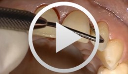
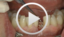
Surgical Treatment of Periodontitis Using a Minimally Invasive Approach
Beck, FrankThis case is an excellent demonstration of the use of the minimally invasive access flap technique for treatment of (chronic) periodontitis in an esthetically critical zone. The access flap was used in conjunction with enamel matrix proteins for regenerative therapy., -
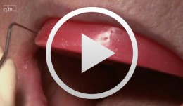
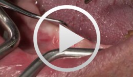
Microsurgical Removal of a Foreign Body from the Mandibular Canal
Schultze-Mosgau, StefanOverview: - Access and incision: Creation of a vestibular pedicled mucoperiosteal flap via a gingival margin incision while preserving the papilla - Removal of vestibular bone in the region of tooth 46 using a microsurgical instrument - Exposure of the neurovascular bundle - Removal of the foreign body - Re-adaptation of the mucoperiosteal flap - Wound closure with atraumatic suture material Contents: Female patient with an indication for microsurgical foreign body removal (removal of a fractured root canal instrument from a previous endodontic treatment of tooth 46) using a surgical microscope. The foreign body extends from the apex into the mandibular canal. -
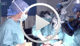
Microsurgical lateral sinus floor elevation (LSFE)
Nölken, RobertOutline: - Incision - Flap mobilization - Lateral sinus fenestration - Elevation of the Schneiderian membrane - Implant bed preparation - Bone chip harvesting at the mandibular angle - Filling of sinus lift lumen with autologous bone chips - Implant insertion - Covering the lateral sinus cavity with collagen membrane - Wound closure List of materials - Zeiss Pro Dent microscope with beam splitter and Panasonic 3 CCD camera - Scalpel holder (Ustomed) with Swann-Morton blades 15C and 12D - Narrow rasp (Hu-Friedy) - Micro-vacuum (Luer Lock Suction Tip, American Dental Systems) - Disposable vacuum tube set (Bexamed) - Disposable draping, Lindau (Aescologic) - Piezosurgery with diamond ball (Mectron) - Microforceps (Hu-Friedy) - Excavator (Martin) - Periodontometer, 1-mm gradation (Hu-Friedy) - OsseoSpeed implant set, Dentsply Implants: Marking drill; Twist drill, 2 mm; Depth gauge; Pilot drill, 2/3.2 mm; Twist drill, 3.2 mm; Tapered drill, 3.2/5 mm; OsseoSpeed TX implant, 5.0 × 11 mm; Closure screw, 4.5/5 mm - Columbia curette (Ustomed) - Micross scraper (Meta) - Needle holder (Ustomed) - Langenbeck wound retractor (Ustomed) - Kelly scissors (Ustomed) - Buchanan endodontic hand plugger (American Dental Systems) - Resorbable collagen membrane (Resodont, Resorba) - Ethilon 5-0 FS-3 (Ethicon) - Prolene 6-0 DA-2 (Ethicon) -
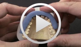
Implant-supported removable restorations in the edentulous jaw
Wolfart, Stefan / Weber, VolkerOutline: - Patient presentation, impression, comprehensive jaw relation records - Wax-up, Fabrication of the provisional restoration - Fabrication of a DVT based planning and drilling template - Surgical procedures for inserting four implants in the mandible - Suturing and relining of the existing denture as a provisional restoration - After 12 weeks: Reentry and insertion of healing abutments - Harvesting a free gingiva graft to extend the attached gingiva - Verifying implant stability using Periotest - Reworking the existing denture to fit on the healing abutments - Impressioning with custom tray (pick-up technique) - Demonstrating the line finder to transfer face axis - Fabricating the three restorations with Locator® attachments, electroplated double crown, precision-milled bar - The matrix and retention parts of the Locator® system, transferring the Locator® abutments to the implants - Fabricating the electroplated copings, intraoral adhesively connecting the electroplated copings to the cast framework (passive fit), finishing and delivery - Removable restoration on a custom-milled bar, clinical and laboratory workflow, delivery - Maintaining implant-supported restorations -
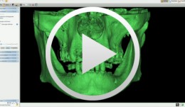
3D planning and template-guided implant insertion in the edentulous jaw
Kirsch, Axel / Ackermann, Karl-Ludwig / Neuendorff, GerhardContents: - Surgical procedures for anchoring a diagnostic guide - Inserting four provisional implants - Impression and bite registration, fabrication of the master cast - Tooth-setup for the temporary restoration - Registering the setup in a silicone index - Duplicating the setup in radiopaque resin for CT imaging - Implant planning using a 3D record of the CT image - Fabricating a transfer template based on the CAD treatment plan using the CAMLOG® Guide System - Inserting the guiding sleeves into the template - Fabricating the final restoration prior to inserting the implants - Vario SR abutments with Vario SR titanium copings for a passive fit - Fabricating a cast titanium framework to reinforce the restoration - Surgical procedures demonstrating the definitive implant insertion - Insertion of six implants for immediate loading - Providing a controlled-clearance fit between the implants and the denture base -
3D planning and template-guided implant insertion in the partially edentulous jaw
Kirsch, Axel / Ackermann, Karl-Ludwig / Neuendorff, GerhardContents: - Planning and preparing a transfer stent - Introduction to on-screen 3D planning - CAD-designed transfer stent - Integrating the CAMLOG® Guide guiding sleeves - Fabricating a therapeutic restoration - Positioning the lab analogs on the master cast using the transfer stent - Titanium bridge frameworks for the temporary restoration - Surgical procedures: implant placement with the transfer template - Hydrocolloid impression taking and bite registration - Temporary insertion of the therapeutic provisional, demonstrating of a passive fit - Definitive CAMLOG abutments - Scanning the abutments, designing the crowns on-screen, machine driven milling of the zirconia crowns, veneering - Preparation for the delivery of the definitive restoration - Connecting the abutments, checking the torque, cementing the crowns, functional checks - Evaluating the completed restoration and assessing the teamwork of the dentist and dental technician -
Immediate placement and all-ceramic restoration in the anterior maxilla - a customized interdisciplinary treatment approach
Happe, Arndt / Nolte, AndreasContents: - Patient presentation and esthetic analysis - Careful extraction of a non-salvageable tooth - Miniplast splint as a surgical template - Harvesting bone from the implant bed - Placing a CONELOG® implant at site 11 - Obtaining a corticospongeous bone cylinder at site 48 - Alveolar augmentation and reconstruction of the buccal bone lamella - Harvesting a connective-tissue graft - Tunneling the vestibular mucosa, various suturing techniques - Insertion of the provisional restorations - 3 months later: Preparing, impression and arbitrary transfer with a bite fork and facebow, temporary restoration - Master cast, new wax-up, determine the emergence profile - Fabricating a hybrid abutment, Scanning the custom abutment, on-screen crown design - Fabricating a zirconia abutment and a feldspathic ceramic veneer - Conditioning and adhesive attachment of the components, final intraoral check - Try in and adhesive cementation -
Implant-supported removable restorations in the edentulous jaw
Wolfart, Stefan / Weber, VolkerContents: - Patient presentation, impression, comprehensive jaw relation records - Wax-up, Fabrication of the provisional restoration - Fabrication of a DVT based planning and drilling template - Surgical procedures for inserting four implants in the mandible - Suturing and relining of the existing denture as a provisional restoration - After 12 weeks: Reentry and insertion of healing abutments - Harvesting a free gingiva graft to extend the attached gingiva - Verifying implant stability using Periotest - Reworking the existing denture to fit on the healing abutments - Impressioning with custom tray (pick-up technique) - Demonstrating the line finder to transfer face axis - Fabricating the three restorations with Locator® attachments, electroplated double crown, precision-milled bar - The matrix and retention parts of the Locator® system, transferring the Locator® abutments to the implants - Fabricating the electroplated copings, intraoral adhesively connecting the electroplated copings to the cast framework (passive fit), finishing and delivery - Removable restoration on a custom-milled bar, clinical and laboratory workflow, delivery - Maintaining implant-supported restorations -
Innovative CAD/CAM treatment approaches for implant-supported fixed restorations
Beuer, Florian / Stimmelmayr, Michael / Schweiger, JosefContents: - Patient presentation - Preparing the implant bed, implant placement, checking implant positions - Securing the insertion posts to the index for fabrication of the cast - Suturing details - Delivery of the adapted long-term provisional - Fabricating the cast and the gingival mask, transferring the pontic emergence profiles to the gingival mask, mask adaptation - The master cast under the strip scanner with scan bodies on the laboratory analogs - CAD crown design and virtual anatomic shaping - CAM fabrication of a zirconia abutment - Adhesively connecting the zirconia abutment to the titanium base - Reentry, split-thickness flap, vestibuloplasty, connecting the zirconia abutments to the implants - Mucosal graft to restore a soft-tissue defect - Intraoral impression of the abutments - Fabricating the definitive lithium disilicate crowns: virtual crown design; CAM milling, characterization of the crowns - Delivery, final adjustments, presentation of the treatment outcome -
Immediate placement and all-ceramic restoration in the anterior maxilla - a customized interdisciplinary treatment approach - Clinical procedure
Happe, ArndtContents: - Patient presentation and esthetic analysis - Careful extraction of a non-salvageable tooth - Miniplast splint as a surgical template - Harvesting bone from the implant bed - Placing a CONELOG® implant at site 11 - Obtaining a corticospongeous bone cylinder at site 48 - Alveolar augmentation and reconstruction of the buccal bone lamella - Harvesting a connective-tissue graft - Tunneling the vestibular mucosa, various suturing techniques - Insertion of the provisional restorations - 3 months later: Preparing, impression and arbitrary transfer with a bite fork and facebow, temporary restoration - Master cast, new wax-up, determine the emergence profile - Fabricating a hybrid abutment, Scanning the custom abutment, on-screen crown design - Fabricating a zirconia abutment and a feldspathic ceramic veneer - Conditioning and adhesive attachment of the components, final intraoral check - Try in and adhesive cementation -