


-
- English
- Deutsch
- Spanish
-
- General Diagnostics
- Treatment
- Tools/Materials
- Basic/General Knowledge
- Research
- Events
- Organisations
-
-
-
-
-
-

-
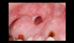

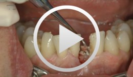
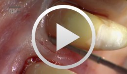
Total reconstruction after closure of maxillary sinus fistula
Dr. Thomas TruningerBefore starting the full-mouth reconstruction, the oral-maxillary sinus fistula (oroantral fistula) has to be closed. -
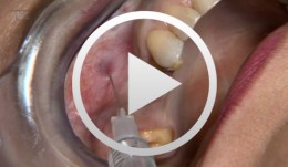
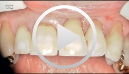
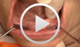
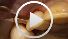
Innovative CAD/CAM treatment approaches for implant-supported fixed restorations
Beuer, Florian / Stimmelmayr, Michael / Schweiger, JosefOutline: - Patient presentation - Preparing the implant bed, implant placement, checking implant positions - Securing the insertion posts to the index for fabrication of the cast - Suturing details - Delivery of the adapted long-term provisional - Fabricating the cast and the gingival mask, transferring the pontic emergence profiles to the gingival mask, mask adaptation - The master cast under the strip scanner with scan bodies on the laboratory analogs - CAD crown design and virtual anatomic shaping - CAM fabrication of a zirconia abutment - Adhesively connecting the zirconia abutment to the titanium base - Reentry, split-thickness flap, vestibuloplasty, connecting the zirconia abutments to the implants - Mucosal graft to restore a soft-tissue defect - Intraoral impression of the abutments - Fabricating the definitive lithium disilicate crowns: virtual crown design; CAM milling, characterization of the crowns - Delivery, final adjustments, presentation of the treatment outcome -
-
SOS - An innovative method for the implantological rehabilitation of the edentulous mandible
Sliwowski, Christoph T. -
-
Regenerative Measures for Osseous Defect Repair and Optimal Esthetics
Sculean, AntonProcedure: Theoretical Part: - Adult male with a deep and broad intraosseous bone defect located on tooth #13 - The indication for modified papilla preservation in the scope of regenerative therapy was established based on the width of the diastema - Regenerative periodontal therapy with Emdogain and a Bio-Oss® cancellous bone graft - Emdogain is applied to the root surface to stimulate regeneration of periodontal structures - To prevent graft collapse and to minimize the risk of development of too large a recession in this esthetically important region, the defect was filled with Bio-Oss® cancellous bone material Practical Part: - The papilla preservation technique was performed using microsurgical instruments - The root surface area was conditioned with 24% EDTA for ca. 2 minutes - Emdogain was applied to the root surface - The defect was filled with the Emdogain/Bio-Oss® mixture - The wound was closed with two mattress sutures one horizontal mattress suture to secure the graft in place, and a second modified vertical mattress suture to tightly close the papilla - A 5-0 suture was used for the horizontal mattress suture, and a 6-0 monofilament was used for the vertical mattress suture - Postoperative care entailed rinsing the wound twice daily for 4 weeks with 0.2% chlorhexidine and ibuprofen analgesia on the first few days after surgery Contents: The patient's jaw displayed a generalized loss of clinical attachment and alveolar bone. His general history was unremarkable; the patient was a non-smoker. Microbiological tests showed large numbers of Actinobacillus actinomycetemcomitans and Porphyromonas gingivalis. The diagnosis was "generalized aggressive periodontitis". After four months of initial therapy consisting of antibiotic combination therapy (amoxicillin + metronidazole), intraoral radiographs showed a deep and wide intraosseous bone defect located mesial and palatal to tooth #13. To preserve this strategically important tooth we opted to perform regenerative therapy with Emdogain and Bio-Oss cancellous bone material. Ten months after regenerative periodontal therapy, the probing depth had decreased by 7 mm, and 5-6 mm of clinical attachment had been gained. At this time, the probing depth was 2-3 mm and intraoral radiographs showed near-complete filling of the osseous defect. -
Regenerative Procedures for Optimized Esthetics at Tooth 11
Schlee, MarkusContents: - Exploration - Incision and Flap Mobilization - Palatal Flap Preservation with Interdental Tissue Preservation - Detoxification and Concrement Removal at 11 - Harvesting of Autogenous Bone Chips from the Spina Nasalis - Conditioning of the Root Surface with EDTA-Gel - Application of Emdogain and Filling of the Bone Defect - Wound Closure Synopsis After Finishing the Initial Treatment for Aggressive Periodontitis, Regenerative Treatment of a Tunnel-Shaped Pocket at Tooth 11 was attempted. Rotation and Crowding of the Buccally Inclined Tooth represented a favorable Etiological Factor. The patient did not wish to receive Orthodontic Treatment to eliminate this Causal Factor after Completion of Primary Treatment. Treatment was therefore limited to the Surgical Regeneration Attempt. The Interdental Space was larger than 3 mm and the Bone Pocket was a mostly Three-Walled Structure, so the Chances of Success were considered to be good. Exploration was first performed to identify the Course of the Defect Margins. Exact knowledge of the Bone Anatomy in all three Planes is essential to successful Incision Planning. A Tunnel-Shaped Defect delimited by Bone in the Region of Tooth 11 with good chances of Periodontal Regeneration was found. A major Challenge of this Procedure is the need to keep the Defect completely covered with Soft Tissue throughout the Healing Process. Cortellini's Papilla Preservation Technique was used for this Purpose. After Incision and Flap Mobilization, it became evident that the Defect only had two Walls in the Coronal Region and that Bone was lacking in the Buccal Region. According to the current Data on Periodontal Regeneration, the Attachment Gain achieved using an Enamel Matrix Protein (Emdogain®) alone can be just as good as that achieved using Emdogain and Bone Graft Material combined. Still, we elected to use a Combination Technique in the Present Case because it provides better Papillary Support. The Graft Material consisted of Autogenous Bone Chips from the Spina Nasalis, which can easily be harvested by Means of the Piezo Technique After gentle Detoxification, the Root Surface was treated with Emdogain. The Defect was then filled with Autogenous Bone Chips and closed by Microsurgical Suture Techniques. Six months after Surgery, Partial Regeneration of the Papilla can be seen. -
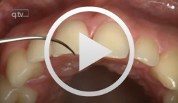
Flap designs for Interdental Tissue Preservation in Periodontal Therapy
Salvi, Giovanni E.Procedure: - Introduction: History -Taking, Examination, Diagnosis, Etiology, Prognosis for Individual Teeth - Four - Phase Treatment Sequence - Modified Papilla Preservation Technique(MPPT) - Simplified Papilla Preservation Technique (SPPT) - Findings 6 months after Surgery Synopsis The Modified and Simplified Papilla Preservation Techniques for conservation of interproximal papillary tissue were designed to provide access to deep and narrow bony defects to enable regenerative periodontal treatment. The Modified Papilla Preservation Technique (MPPT) was designed to ensure tension-free primary closure via barrier membranes in patients with small interdental spaces. The Simplified Papilla Preservation Technique(SPPT) is used to gain access to narrow interdental spaces ( < 2mm) and to deep defects in the lateral tooth region. Apart from preserving primary wound closure in the interdental space, the two techniques also serve to keep the membrane from collapsing into the bony defect. Both MPPT and SPPT employ special suture techniques to ensure tension-free primary closure of the interdental space. This video clip also serves to demonstrate that the two techniques of interproximal tissue preservation can also be used for periodontal interventions without regenerative measures. -
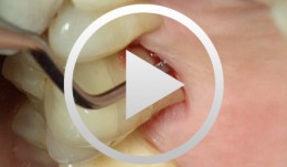
The Carnevale Technique: Hemisection/Trisection of Molars with Furcation Involvement
Hürzeler, Markus B.Procedure: - Apical Flap Repositioning - Trisection of the Upper Molar - Extraction of the Distobuccal Root - Tangential Preparation of the Abutment Teeth - Temporary Relining Contents: Molars with furcation involvement have a shorter long-term prognosis for tooth retention than single-rooted teeth. Apart from replacing these teeth with implants, they can also be treated by hemisection or trisection with the goal of eliminating furcation and creating single-root conditions. Studies on the long-term stability of teeth treated by hemisection / trisection show mixed results. Some investigators have found failure rates of up to 40 percent. In Gianfranco Carnevale's group, on the other hand, success rates of more than 90 percent have been reported for a 10-year follow-up period. Pretreatment: Initial work on the abutment teeth, which had grade II-III furcation involvement, was done six to eight weeks after conservative periodontal therapy in preparation for placement of the long-term temporary. Preparation was done tangentially, up to the bone level, to spare as much dental hard tissue as possible while eliminating all recesses and root concavities. A metal-reinforced long-term temporary was used to splint the prepared teeth. Endodontic treatment of the affected teeth was subsequently performed. Surgical procedure: Apical displacement of the gingiva around the involved teeth was done before incision. A mucosal fl ap was created on the buccal and palatal sides. Trisection of the teeth was then performed. After dissecting and excising the distobuccal root, intraoperative preparation of the abutment teeth was carried out. Temporary relining was another important step, the objective of which was to splint the root and to prevent tilting. Further treatment: Impressions for the defi nitive restoration were made six months postoperative. The restoration margins of the tangential preparation were defined using a master model. The definitive reconstruction had fine metal margins. -
Gap Closure with a Minor Incisal Edge Restauration
Klaiber, BerndProcedure: - Incisal edge restoration at Tooth 11 - Sketch prior to widening procedure in anterior teeth region - Placement and shaping of matrix band retainer used for tooth widening - Application of composite material and spreading with small Heidemann spatula to establish stable and broad approximal contact - Application of dentin and enamel composite - Shaping and completing; positioning of the lateral edge lines; shaping the interincisal triangle. - Creation of an invisible transition, composite/enamel, with scalpel #15 - Polishing Materials Adhesive: Optibond Fl (Kerr) Composite: Enamel HFO (Micerium) Dentin UD4, UD3,5 and UD3 Enamel GE2 Opalescence OBN Flowable Composite: Tetric Flow A4 (Vivadent) Temporary Composite for shaping of matrix band retainer SystepOnlay (Vivadent) -
PSI - the Periodontal Screening Index
Belz, Dieter -
Mandibular Distraction Osteotomy
Schleier, Peter / Schultze-Mosgau, StefanProcedure - Indications and preoperative planning - Incision technique and osteotomy - Placement of distraction apparatus - Wound closure and postoperative regimen Materials: V2-Alveolar distractor, Medartis (Switzerland) 2,0mm Screws for Osteosynthesis, Medartis (Switzerland) Vicryl Suture, Ethilon (USA)