


-
- English
- Deutsch
- Spanish
-
- General Diagnostics
- Treatment
- Tools/Materials
- Basic/General Knowledge
- Research
- Events
- Organisations
-
-
-
-
-
-

-
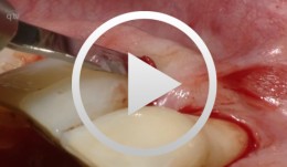

Flap surgery using a modified papilla preservation technique
Salvi, Giovanni E.Outline - Introduction: Patient history, clinical findings, diagnoses, etiology, prognosis of individual teeth - Treatment planning for phases - Modified papilla preservation technique (MPPT) - Simplified papilla preservation technique (SPPT) -Clinical situation 6 months postoperatively -
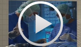
Operative Therapiekonzepte zur Entfernung retinierter unterer Weisheitszähne
Schultze-Mosgau, Stefan / Neukam, Friedrich Wilhelm / Basting, GerdGliederung: - Schematische und röntgenologische Demonstration unterschiedlicher Retentionsformen unterer, retinierter Weisheitszähne, Darstellung der Indikation zur Entfernung - Erstellung der Behandlungsunterlagen, Aufklärungsgespräche mit Darstellung der Komplikationen - Demonstration des operativen Vorgehens: Lokalanästhesie, Schnittführung, Schutz des N. lingualis, Osteotomie, Nahttechnik. Die Entfernung unterer, retinierter Weisheitszähne gehört zu den häufigsten dento-alveolär-chirurgischen Eingriffen. Durch die enge anatomische Lagebeziehung zu den benachbarten Zähnen und dem N. alveolaris inferior besteht bei der operativen Entfernung die Gefahr einer Schädigung der umgebenden Strukturen. Die Kenntnis der verschiedenen Retentionsformen und eine geeignete, atraumatische Operationstechnik ist für eine komplikationslose Entfernung von Bedeutung. Nach der Leitungsanästhesie des N. alveolaris inferior und des N. buccalis wird die Schnittführung so gewählt, dass ein vestibulär gestielter Mukoperiostlappen gehoben werden kann. Nach dem lingualen, subperiostalen Einführen eines Raspatoriums zum Schutz des N. lingualis wird durch die bukkale Osteomie mit kugelförmigen Hartmetallfräsen der Weisheitszahn bis zu seiner größten Zirkumferenz freigelegt und durch vorsichtige Luxationsbewegungen entfernt. -

-
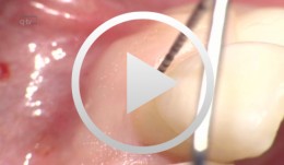
Periodontal Preserve Therapy (Examples)
Clotten, StefanContent: - Periodontal maintenance therapy for teeth 34 and 35, including the regeneration of a bone defect using bone replacement material, collagen membrane and sutures. - Curettage for treatment of periodontal pockets. - Treatment of gingival pressure sores caused by tight-fitting orthodontic apparatus. - Incision of buccal attachment to relieve gingival pressure for elimination of gingival recession. -
Errors and Checklists
Franck Renouard -
Tendency of skeletal class 3 and a missing front tooth
Dr. Dominik BüchiIn this clinical case session you are going to learn how to plan and execute treatment in a partially edentulous patient situation applying implants within the overall reconstructive treatment concept. In particular you will learn the importance of the preparatory phases of therapy and to plan and treat with the prosthetic aim in mind. -
Periodontitis case, fixed reconstructions on teeth, implants and tooth-implant combination
Prof. Niklaus Lang and B. RöthlisbergerPeriodontitis case with acute pain and furcation involvement, fixed reconstructions on teeth, implants and tooth-implant combination. -
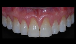
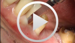
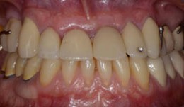
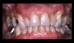
Connective tissue graft with tunnel technique for root coverage
Ignacio Sanz MartinRecessions in the anterior maxillary sextant due to traumatic tooth brushing. Connective Tissue graft with tunnel technique for root coverage. -
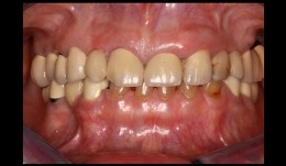
Esthetic and functional dental rehabilitation
Dr. Dominik BüchiMale patient (*1955) treated by Dr. D. Büchi. Functional and esthetic rehabilitation through replacement of insufficient crowns and bridges. Additionally implants were placed in the posterior part, to maximize the masticatory performance. -
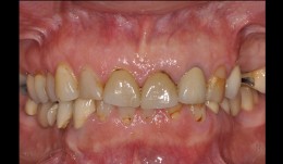
Esthetic and functional rehabilitation
Sven Mühlemann58-year-old woman desires a total refurbishment by reason of compromised esthetics of her front teeth and missing teeth in the upper left jaw. In addition the bridges in the mandibular left and right show chippings. -
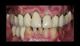
Esthetic and functional restoration
Thomas HitzThe old restorations showed several ceramic fractures and in some areas the framework was exposed. The patient's main concern was the unesthetic appearance of the anterior crowns in the maxilla due to these chippings. -
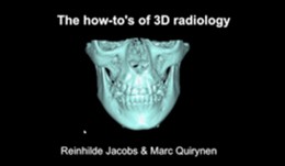
Essentials of 3D radiology
Prof. Reinhilde Jacobs and Prof. Marc QuirynenCone beam computer tomography (CBCT) has become an important tool for diagnostics and planning in implant dentistry. This module explains you all the important aspects of the CBCT.