


-
- English
- Deutsch
- Spanish
-
- General Diagnostics
- Treatment
- Tools/Materials
- Basic/General Knowledge
- Research
- Events
- Organisations
-
-
-
-
-
-

-

Flap designs for Interdental Tissue Preservation in Periodontal Therapy
Salvi, Giovanni E.Procedure: - Introduction: History -Taking, Examination, Diagnosis, Etiology, Prognosis for Individual Teeth - Four - Phase Treatment Sequence - Modified Papilla Preservation Technique(MPPT) - Simplified Papilla Preservation Technique (SPPT) - Findings 6 months after Surgery Synopsis The Modified and Simplified Papilla Preservation Techniques for conservation of interproximal papillary tissue were designed to provide access to deep and narrow bony defects to enable regenerative periodontal treatment. The Modified Papilla Preservation Technique (MPPT) was designed to ensure tension-free primary closure via barrier membranes in patients with small interdental spaces. The Simplified Papilla Preservation Technique(SPPT) is used to gain access to narrow interdental spaces ( < 2mm) and to deep defects in the lateral tooth region. Apart from preserving primary wound closure in the interdental space, the two techniques also serve to keep the membrane from collapsing into the bony defect. Both MPPT and SPPT employ special suture techniques to ensure tension-free primary closure of the interdental space. This video clip also serves to demonstrate that the two techniques of interproximal tissue preservation can also be used for periodontal interventions without regenerative measures. -
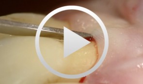
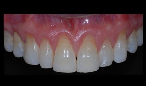
Regenerative Procedures for Optimized Esthetics at Tooth 11
Schlee, MarkusContents: - Exploration - Incision and Flap Mobilization - Palatal Flap Preservation with Interdental Tissue Preservation - Detoxification and Concrement Removal at 11 - Harvesting of Autogenous Bone Chips from the Spina Nasalis - Conditioning of the Root Surface with EDTA-Gel - Application of Emdogain and Filling of the Bone Defect - Wound Closure Synopsis After Finishing the Initial Treatment for Aggressive Periodontitis, Regenerative Treatment of a Tunnel-Shaped Pocket at Tooth 11 was attempted. Rotation and Crowding of the Buccally Inclined Tooth represented a favorable Etiological Factor. The patient did not wish to receive Orthodontic Treatment to eliminate this Causal Factor after Completion of Primary Treatment. Treatment was therefore limited to the Surgical Regeneration Attempt. The Interdental Space was larger than 3 mm and the Bone Pocket was a mostly Three-Walled Structure, so the Chances of Success were considered to be good. Exploration was first performed to identify the Course of the Defect Margins. Exact knowledge of the Bone Anatomy in all three Planes is essential to successful Incision Planning. A Tunnel-Shaped Defect delimited by Bone in the Region of Tooth 11 with good chances of Periodontal Regeneration was found. A major Challenge of this Procedure is the need to keep the Defect completely covered with Soft Tissue throughout the Healing Process. Cortellini's Papilla Preservation Technique was used for this Purpose. After Incision and Flap Mobilization, it became evident that the Defect only had two Walls in the Coronal Region and that Bone was lacking in the Buccal Region. According to the current Data on Periodontal Regeneration, the Attachment Gain achieved using an Enamel Matrix Protein (Emdogain®) alone can be just as good as that achieved using Emdogain and Bone Graft Material combined. Still, we elected to use a Combination Technique in the Present Case because it provides better Papillary Support. The Graft Material consisted of Autogenous Bone Chips from the Spina Nasalis, which can easily be harvested by Means of the Piezo Technique After gentle Detoxification, the Root Surface was treated with Emdogain. The Defect was then filled with Autogenous Bone Chips and closed by Microsurgical Suture Techniques. Six months after Surgery, Partial Regeneration of the Papilla can be seen. -
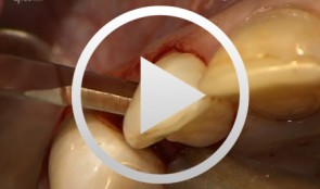
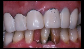
Regenerative Measures for Osseous Defect Repair and Optimal Esthetics
Sculean, AntonProcedure: Theoretical Part: - Adult male with a deep and broad intraosseous bone defect located on tooth #13 - The indication for modified papilla preservation in the scope of regenerative therapy was established based on the width of the diastema - Regenerative periodontal therapy with Emdogain and a Bio-Oss® cancellous bone graft - Emdogain is applied to the root surface to stimulate regeneration of periodontal structures - To prevent graft collapse and to minimize the risk of development of too large a recession in this esthetically important region, the defect was filled with Bio-Oss® cancellous bone material Practical Part: - The papilla preservation technique was performed using microsurgical instruments - The root surface area was conditioned with 24% EDTA for ca. 2 minutes - Emdogain was applied to the root surface - The defect was filled with the Emdogain/Bio-Oss® mixture - The wound was closed with two mattress sutures one horizontal mattress suture to secure the graft in place, and a second modified vertical mattress suture to tightly close the papilla - A 5-0 suture was used for the horizontal mattress suture, and a 6-0 monofilament was used for the vertical mattress suture - Postoperative care entailed rinsing the wound twice daily for 4 weeks with 0.2% chlorhexidine and ibuprofen analgesia on the first few days after surgery Contents: The patient's jaw displayed a generalized loss of clinical attachment and alveolar bone. His general history was unremarkable; the patient was a non-smoker. Microbiological tests showed large numbers of Actinobacillus actinomycetemcomitans and Porphyromonas gingivalis. The diagnosis was "generalized aggressive periodontitis". After four months of initial therapy consisting of antibiotic combination therapy (amoxicillin + metronidazole), intraoral radiographs showed a deep and wide intraosseous bone defect located mesial and palatal to tooth #13. To preserve this strategically important tooth we opted to perform regenerative therapy with Emdogain and Bio-Oss cancellous bone material. Ten months after regenerative periodontal therapy, the probing depth had decreased by 7 mm, and 5-6 mm of clinical attachment had been gained. At this time, the probing depth was 2-3 mm and intraoral radiographs showed near-complete filling of the osseous defect. -
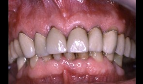
Periodontitis case, fixed reconstructions on implants (bridge and single crown)
Dr. Bjarni E. Pjetursson and Niklaus LangMale patient *1953 by B. E. Pjetursson, N. P. Lang (2001-2003). Periodontitis case, fixed reconstructions on implants (bridges in the mandible and single crown in the maxilla). -
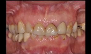
Patient referred has reconstruction in maxilla, shortened dental arch reconstructed
Prof. Niklaus Lang and M. LulicMale patient *1941 by M. Lulic, N. P. Lang (2005-2007). Patient referred has reconstruction in the maxilla, former smoker, shortened dental arch reconstructed with 3 single crowns on implants. -
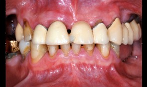
Old reconstructions and missing teeth - a comprehensive case
Dr. Christian LeutertMissing posterior teeth and old unesthetic reconstructions - see how this case was solved by Dr. Christian Leutert at the University of Zurich. -
Esthetic and functional rehabilitation
Sven Mühlemann58-year-old woman desires a total refurbishment by reason of compromised esthetics of her front teeth and missing teeth in the upper left jaw. In addition the bridges in the mandibular left and right show chippings. -
Esthetic and functional dental rehabilitation
Dr. Dominik BüchiMale patient (*1955) treated by Dr. D. Büchi. Functional and esthetic rehabilitation through replacement of insufficient crowns and bridges. Additionally implants were placed in the posterior part, to maximize the masticatory performance. -
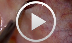
Connective tissue graft with tunnel technique for root coverage
Ignacio Sanz MartinRecessions in the anterior maxillary sextant due to traumatic tooth brushing. Connective Tissue graft with tunnel technique for root coverage. -
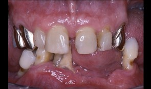
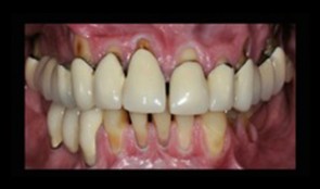
Comprehensive treatment of periodontitis with full mouth reconstruction
B. E. Pjetursson, L. Adriaens, N. P. LangFemale patient *1942 by B. E. Pjetursson, L. Adriaens, N. P. Lang (2004-2006). Comprehensive treatment of periodontitis with full mouth reconstruction.