


-
- English
- Deutsch
- Spanish
-
- General Diagnostics
- Treatment
- Tools/Materials
- Basic/General Knowledge
- Research
- Events
- Organisations
-
-
-
-
-
-

-

THE BOX - THE GLOBAL OSTEOLOGY COMMUNITY PLATFORM
It is a big challenge to link scientists and practitioners in a continuously growing global context and also a fundamental part of the Osteology Foundation’s mission: promoting research and education in oral tissue regeneration worldwide. With this in mind, the Osteology Foundation has set up the novel Global Osteology Community Platform THE BOX, which on the one hand provides information and tools, but on the other also connects scientists and practitioners worldwide and supports all existing activities of the Osteology Foundation online. -
Learning from the Experts – Research Academy Lucerne
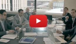
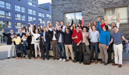
Research drives advances in science - this is why the Osteology Foundation is committed to running an intensive course on good scientific practice and research methodology. 26 participants from 12 different countries attended the Core Module in Lucerne this year. The one-week intensive course provided an overview of the planning and running of a study project from A to Z. In classroom sessions with leading experts, such as Niklaus Lang and Reinhard Gruber, case studies and exercises in teams the young researchers learned the Essentialsof Research Methodology, how to plan, conduct, evaluate and publish the results. The participants thereby particularly liked the highly interactive classes and the casual atmosphere. The interaction among the participants as well as the experts was regarded as extremely valuable. They discovered that they have very similar experiences and problems in their research practice despite coming from different countries and cultures from all-over the world, such as Egypt, Brazil, Europe and China. The dates for all up-coming courses can be found on the Osteology Research Academy website. Education Grants are available for participation in the courses. Application is possible online. Deadline: 1 December. I loved the informal atmosphere. The lecturers were engaging and inspiring, and gave me the opportunity to dispel several research doubts I had. Elena Calciolari, United Kingdom An outstanding, well-structured course, with the worlds best experts in the field of dental research. Mahmoud Shalash, Egypt I feel very privileged to have been selected for an education grant from the Osteology Foundation and to be attending the Osteology Research Academy, learning from great renowned professors. This will definitely make a big difference in my career. Vanessa Camillo de Almeida, Brazil (right in picture) The ORA Lucerne gave me the opportunity to meetinteresting colleaguesfrom all over the worldin a great atmosphere. Attending high-level presentations and workshops broadened my scientific horizon. Jonas Lorenz, Germany Attending the Research Academy Lucerne was of greatest value for gaining knowledge on practical aspects on clinical and pre-clinical research provided by experts from different disciplines. The great course environment provided the opportunity to interactively discuss and learn how to work within a team, especially during the workshops. Aliye Akcali, Turkey An excellent course for young clinicians that are heading for research in the field of oral tissue regeneration. Bo Chen, China -
New Large Clinical Grants Programme
The Osteology Foundation calls for applications for the new Large Clinical Grants. Theprogram aims to support hypothesis driven clinical research by well-established research groups with demonstrated clinical research expertise. There are two stages in the application process: Stage 1 Applicants can submit an abstract of their proposed project by15 November. The Osteology Science Committee selects projects which then progress to the next stage. Stage 2 Applicants are invited to submit their full application. The submissions are then evaluated for potential funding by the Osteology Science Committee. Study types Research types falling within the scope of the Osteology Foundation are for example: RCT (also small multi centres) and quasi RCT Case controlled studies Prospective cohort studies Surgical techniques (also in combination with biologics, drugs and devices) e.g.: Biologics Dugs (also e.g. phase I / phase II - pilot studies, proof of principle) Devices (also e.g. phase I / phase II - pilot studies, proof of principle) Combination products Who can apply? Well-established research groups that demonstrated clinical research expertise. Amount of funding Grants are limited to 350000 Swiss Francs with a maximum project duration of three years. Awarding criteria Hypothesis answering a clinical research question relevant to the field of oral and maxillofacial regeneration Methodology Facilities and expertise to conduct a GCP study Sound and transparent budget Application guidelines Apply now! -

Osteology Researcher Grants - apply now!
The Osteology Foundation offers besides its established Advanced Researcher Grants a special grant program for young researchers: the Osteology Young Researcher Grants, which support researchers in realizing their own research project and fuelling their research career. The Osteology Young Researcher Grants program (new since 2014) is addressed to all clinicians and researchers (primary investigators) currently undergoing post-graduate education or within three years thereafter. The grants are limited to 30000 Swiss Francs with a maximum project duration of one year. The new Young Researcher Program is dedicated to projects with sound hypothesis, and aims that show thinking out of the box. The Osteology Advanced Researcher Grants program is addressed to clinicians and researchers at universities and in private practice (having an university affiliation) conducting research in the field of dental tissue regeneration. It is dedicated to projects with hypothesis and aims focusing on clinical relevance bolstered with preliminary work within a research group setting. In general, grants are limited to 100000 Swiss francs with a maximum project duration of two years. If you have an interesting research project fitting to one of the Osteology Foundation grants programmes you are invited to submit your application before June 15, 2015. Detailed information as well as the application guidelines for the research and education grants are available from the Osteology Website. -
New Osteology Research Scholarship Programme
Research drives advances in science, and promoting research in the field of oral tissue regeneration is one of the core activities of the Osteology Foundation. That is why the Foundation is continuously expanding its activities in this field. New in 2015: the Osteology Foundation is now offering one-year Research Scholarships for young investigators to develop their career in the field of oral tissue regeneration. The aim of the Osteology Foundation is to offer scholars an outstanding environment and expertise on clinical and basic research. Osteology Scholarship Centres The scholars will work on a research project teaming up with their mentors at one of the Osteology Scholarship Centres, which are among the worlds leading research institutes in oral regeneration. The Osteology Scholarship Centres are: University of Gothenburg (Prof. Christer Dahlin) University of Michigan School of Dentistry (Prof. William Giannobile) Medical University of Vienna School of Dentistry (Prof. Reinhard Gruber) University of Zurich (Prof. Christoph Hmmerle) Amount of funding and application details The Research Scholarships consist of 35000 Swiss Francs per scholar for a twelve month stay in one of the Osteology Scholarship Centres, and are offered annually. All young investigators with an academic affiliation can apply. The Research Scholarships are awarded based on the evaluation of a motivation letter, a curriculum vitae and a recommendation letter. Financial needs are also taken into account. Deadline for the submission of applications is May 15, 2015. Applicants will be notified about acceptance by July 15, 2015. Develop your research career in oral regeneration, andapply for an Osteology Research Scholarship now! -

Osteology Researcher Grants: New publications from funded studies
The results of two pre-clinical studies funded by the Osteology Foundation have been published recently in the peer-reviewed journal Clinical Oral Implants Research. The two studies reveal important findings that will help to better understand the mechanisms of bone loss and soft-tissue regeneration. Supporting research into all aspects of oral tissue regeneration is a core task of the Osteology Foundation. Over recent years, the Foundation has supported a number of pre-clinical and clinical studies that have led to about 30 publications in international peer-reviewed journals. In 2014 the results from two pre-clinical studies supported by the Osteology Foundation were published in the high-ranking journal Clinical Oral Implants Research. Both studies provide important insights into basic mechanisms in bone loss and tissue regeneration. Pre-clinical rat model for studies on osteoporosis A study by Xi Ling Liu and co-workers from Hong Kong investigated the skeletal site-specific response to ovariectomy in a rat model (Liu 2014). The investigators examined changes in bone density and microcarchitecture in different bone sites and found significant differences, which is highly relevant for other studies using this animal model for osteoporosis-related studies. After 36 weeks the researchers observed that jaw bones and cranial bones in ovariectomized rats only showed a minor reduction in bone mineral density, whilst long bones, lumber vertebra and ilium in the same animals showed significant bone loss compared to the baseline. A significant deterioration of the trabecular structure was detected in the long bones and vertebra, while jaw bones remained relatively stable. Overall, femur and tibia displayed the largest bone loss. Relevant information for further studies using the model The study by Liu et al. assessed for the first time the systemic site-specific bone loss and microarchitecture changes in ovariectomized rats, a widely used animal model that mimics the estrogen-deficiency-induced bone loss, and one which is approved by the FDA for osteoporosis. The results provide valuable information for the selection of bone sites and observation times in further studies using this animal model for osteoporosis studies a topic that is becoming increasingly important, since osteoporosis, along with the aging of the population, has developed into a relevant health burden. Crosslinked and non-crosslinked collagen matrices A different topic investigated the study by Daniel S. Thoma and his co-workers from Zurich (Thoma 2014). Their study, which was also funded by a Research Grant from the Osteology Foundation, investigated in a rat model whether the crosslinking of a collagen matrix, as well as the addition of recombinant human platelet-derived growth factor-BB (rhPDGF-BB), influenced tissue integration, angiogenesis and degeneration of the collagen matrices. Four treatment modalities were tested in an ectopic model in rats and compared for the study: the cross linked matrix with and without rhPDGF-BB, and the non-cross-linked matrix with and without rhPDGF-BB. In 50 rats, the four combinations were planted in four unconnected pouches on the back of each animal. Descriptive histology and histomorphometric assessments were performed at 2, 4, 8, 16 and 24 weeks after the surgery. Angiogenesis and stability of the collagen matrices The study showed that the compact layer of the non-cross linked matrix delayed angiogenesis and connective tissue formation, while the spongeous cross-linked matrix facilitated early vascularization and also demonstrated network presence over a longer time span. No statistically significant effects of rhPDGF-BB were observed for any of the evaluated parameters. Different characteristics for different indications These results revealed that both types of matrices can be successfully incorporated into the tissue. However, the matrix composition influenced the healing patterns. The study therefore revealed important information on the use of the two different collagen matrices for different indications, since they provide different characteristics due to their different designs. These are important findings that help to evaluate the efficacy of different matrices for different indications and to understand why such differences exist. References Liu XL, Li CL, Lu WW, Cai WX, Zheng LW. Skeletal site-specific response to ovariectomy in a rat model: change in bone density and microarchitecture. Clin Oral Implants Res. 2014 Mar 5. doi: 10.1111/clr.12360. Thoma DS, Nnni N, Benic GI, Weber FE, Hmmerle CH, Jung RE. Effect of platelet-derived growth factor-BB on tissue integration of cross-linked and non-cross-linked collagen matrices in a rat ectopic model. Clin Oral Implants Res 2014 Sep 30. doi: 10.1111/clr.12496. Further information Published studies supported by Osteology Research Grants Information on Osteology Research Grants -
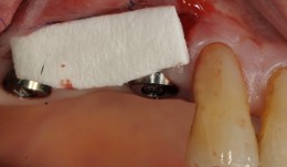
Study Protocols: Soft-Tissue Augmentation
based on the book chapter by Daniel S. Thoma and Ronald E. Jung Summary Soft tissue augmentation using autogenous grafts to restore keratinized tissue volume are described here. Synthetic and biological dermal substitutes, developed initially for treating burns, offer great potential as alternatives to these grafts which have certain limitations (particularly collagen-based products). Two protocols are presented, both for randomized controlled trials. The authors specify two research questions and two sets of defined eligibility criteria, study timelines with relevant clinical steps and time points for measuring clinical endpoints, protocol-specific measures and patient care regimens. Photos are provided of surgical sites before, during and after the procedures. The first study compares autogenous palate grafts (plus an apically positioned flap) with a soft tissue substitute in patients with an implant-supported prosthesis abutted by keratinized mucosa of less than 1 mm thick. Primary outcome measures include keratinized mucosal width, as determined by probing, and parameters relating to safety and effectiveness. There are phases for pre-baseline management, surgery, follow-up of 6 months, and a 5-year assessment of long-term safety, effectiveness and esthetics. The second protocol is for soft tissue volume augmentation in patients requiring volume increase in a single-tooth gap after implant placement. The positive control is a connective tissue graft, with endpoints including gain in mucosal thickness and esthetics, and a follow-up of 3 months. Open full-text PDF (1.9 MB) -
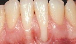
Study Protocols: Root Coverage Regeneration
based on the book chapter byAnton Sculean and Sofia Aroca Summary This protocol relates to root coverage in cases of gingival recession from trauma or disease. Good results can be obtained with a coronally advanced flap technique coupled with connective tissue grafts, but harvesting the tissue is associated with negative factors. An alternative method is soft tissue grafting as used by this protocol, which compares methods and esthetic outcomes in patients with troublesome single and multiple recessions. The selection criteria are stated clearly for patients with Miller class I, II or III gingival recessions in the maxillary or mandibular arch with apicocoronal extension and a recession depth of more than 2 mm. Recruitment is on a multicenter basis, with stratification and randomization into test and control groups, and restrictions on revealing the type of intervention to the surgeons. There are photographs of test and control sites pre-, intra- and postoperatively and at 12 months. Weekly evaluations are carried out for the first month, with a total of 14 visits over the first year. Follow-up is for 35 years. Percentage root coverage is the primary endpoint, with secondary endpoints including patient-reported pain, root sensitivity, tissue thickness and long-term stability. Potential adverse events are listed together with a grading system to assess them. The authors stress the need for surgeon learning curves and calibration of examiners, techniques and equipment, blinded procedures, and engagement of a statistician. Open full-text PDF (1.4 MB) -
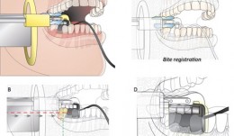
Analytical Methods: Imaging
based on the book chapter by Erika Benavides and Hector F. Rios Summary Clinical imaging allows qualitative and quantitative assessment of hard and soft oral tissues in patients after procedures such as ridge augmentation. This comprehensive guide deals with in vivo and ex vivo and intraoral and extraoral methods. It covers standardized digital intraoral radiography, cone beam (CBCT) and multislice (MSCT) computed tomography, subtraction radiography, ultrasound, optical coherence tomography (OCT), and newer methods such as CAD/CAM (computer-aided design/computer-aided manufacturing) and fractal analysis. There are descriptions on how each modality words, and outlines of their common applications, relative merits and limitations. Among the issues addressed are wave type (e.g infrared versus ionizing radiation), signal noise, distortion, mage resolution, field of view, scanning time, patient trauma and artifacts such as beam-hardening. Illustrations are used throughout to clarify the discussion. The authors point out which methods are preferable for macrostructure, and which for microstructure; for mineralized tissues, and which for soft tissue contours. Some are better for revealing the functions and dynamics of tissues or volumetric changes; some are better at 3-D visualizations and complex typography; some involve exposing patients to ionizing radiation. The authors stress the importance of integrating information from different modalities (such as CAD/CAM with CBCT) and from different time points, to optimize information available for interpretation. Open full-text PDF (1.8 MB) -
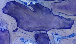
Analytical Methods: Histology
based on the book chapter by Dieter D. Bosshardt Summary Histological assessment, which can be quantitative or qualitative, is an indispensable tool for investigation of soft tissue regeneration. Harvesting tissues raises ethical considerations, but less invasive forms of evaluation, such as clinical measures and imaging, only complement the descriptive analysis, rather than replace it, by showing how cells respond to biomaterials in vivo. This chapter explores various methods for evaluating hard and soft oral tissues, such as histomorphometry, in situ hybridization and autoradiography, in the context of procedures like socket preservation, ridge augmentation and sinus floor elevation. As well as outlining suitable endpoints and landmarks, it discusses tools and techniques for taking biopsies, cutting, sectioning and embedding samples, and the use of microscopy and illumination. The authors address the challenges posed by different biomaterials -- not all can be sectioned, and some react with the chemicals used for processing samples. Thus tissue sections may be of unpredictable quality and be affected by artifacts introduced before fixation (if the tissue gets crushed during harvesting or contaminated by oral pathogens) or during fixation or preserving. Other challenges include the acquisition, processing, analysis and storage of digital images, and the huge variations in analytical equipment and methods used in different laboratories, where the need to apply laboratory standards and minimize examiner variation is emphasized. Open full-text PDF (1.9 MB) -

Statistical Analysis
based on the book chapter by Jrg Hsler Summary This chapter will enable clinical investigators to select statistical methods that extract results and conclusions from quantitative data which are both meaningful and can be compared with findings of other studies. Among the key issues discussed are the need to develop a data analysis plan, to engage a biostatistician early in the design process, and to conduct a power analysis to determine how many participants should be recruited and how many to include in treatment and control groups. There are explanations of different types of randomization processes (simple, stratified, balanced or computer-generated), error probabilities and error corrections, data distribution, and rejection of the null hypothesis on the basis of a statistical test decision. The authors explain how to choose suitable endpoints and analytical tools for specific clinical scenarios that relate to the study hypothesis. The role of descriptive measures, statistical estimates and graphs is made clear, including the concepts of confidence intervals, means, medians and standard deviations. They also describe how statistical models such as analysis of variance (ANOVA) are used to explore relations between variables, and one-way and two-way tests are used to explore relations between impact factors. The role of multiple testing is clarified, and the use of Tukey and Bonferroni corrections, Newman--Keulbs, Scehhe and Dunn devices, and Fishers least significant device (LSD) in error correction. Open full-text PDF (1.1 MB) -
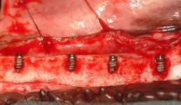
Horizontal Ridge Augmentation
Ridge augmentation is sometimes needed to establish proper soft and hard tissue support before placing a dental implant. The horizontal augmentation procedure is commonly used in dental clinics to restore bone defects in people with destructive gum disease. There are many variables to investigate to optimize these treatments, for which several animal models exist. Defects can be created surgically in skeletally mature animals to study changes over time. The site of both defects and implants can also be varied, and the merits of staged or simultaneous tooth extraction and insertion of implants can be evaluated. Different types of graft material (block, particulate or autogenous), bone fillers and bone substitutes can be applied, and resorbable and non-resorbable, natural and synthetic membranes can also be compared. A major aim of these studies is to identify procedures and materials giving the most predictable outcomes. The chapter describes valid endpoints evaluated by histomorphometry, specific staining techniques, and a host of imaging modalities. It also covers other aspects of successful, reproducible experiments, such as planning, report writing, ethical approval, funding applications and submission, with the overall aim of producing new, safe clinical innovations that rapidly restore quality of life to patients.