


-
- English
- Deutsch
- Spanish
-
- General Diagnostics
- Treatment
- Tools/Materials
- Basic/General Knowledge
- Research
- Events
- Organisations
-
-
-
-
-
-

-

Ridge Preservation
The alveolar ridge is the bony tissue that surrounds a fully erupted tooth. Its structure may be compromised after extraction of the tooth, but it can be preserved by use of bone substitutes, dental implants and buccal overbuilding with soft tissue grafts. Ridge preservation techniques must be developed in animals before clinical trials in patients can be conducted, and various processes, such as wound healing, can be explored that are not feasible to study in people. Dogs are most commonly used because of their similar tooth types, root structures and fast remodeling time. Several preclinical studies are described in this chapter – mostly using the dog mandible – that compare different experimental conditions, biomaterials, grafts and implants after extraction of teeth. These provide information on the timing and sequence of extraction and grafting procedures, and timepoints for assessing tissue shrinkage and resorption of alveolar bone. Other studies address osseous resorption of flaps, the lack of a periodontal ligament, buccal overbuilding, alveolar plate resorption and inflammatory reactions. Jaw casts, morphometry, histology and high-resolution imaging are used as valid endpoints relating to the alveolar process and socket walls, and the cellular content and mineralization of tissues. The protocols outlined in this chapter will contribute to the accuracy with which preclinical studies predict responses in people. -
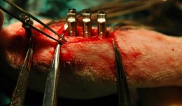
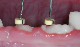
Osseointegration of Implants
The complex process of osseointegration involves formation of an interface between a dental implant and the host’s bone, without intervening soft tissue. Formation depends on qualities of the implant surface, such as its roughness and porosity, as well as characteristics of the host. Titanium coatings offer a good osseointegration surface. After preliminary tests to characterize bone-contacting materials and toxicity reactions, animal models are used to assess different aspects of bone remodeling. This chapter describes several studies for determining the biocompatibility of new implant materials, and their mechanical stability and safety. Models include the tibia of rats and rabbits, and the mandible of the dog, in which different shapes and sizes of implants and fixation techniques can be compared, and the best parameters for quantifying tissue reactions can be established. Mechanical strength testing using measures of torque and shear, for example, help determine how much bone should be in contact with the implant in order to promote osseointegration. The quality of new bone can then be assessed in terms of area, volume and extent of integration. The use of standardized methodology for sample preparation is also emphasized, with detailed description of the Donath technique, which offers many benefits but is expensive, time consuming and requires for specially trained technicians. -
Periodontal Regeneration
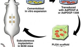
Animals with defects of the gum can be used to investigate the effectiveness and safety of scaffold materials, devices and biologics for bone and soft tissue repair, before they are tested in people. Living models are essential for observing changes in structure and function over time, sometimes long periods, during the remodeling and healing phases. This chapter recommends which animals are best suited for studying agents such as growth factors and barrier membranes. They include both small and large animals, such as rats, dogs and primates, with both natural defects and surgically or ligature-induced defects. The design of the studies is addressed specifically, with instructions on the creation of various standardized defects, and how to care for the animals before and after surgery, including management of biohazardous materials such as viral vectors. The roles of institutional guidelines and the requirements of regulatory bodies and animal housing authorities are also covered. Investigators who are studying the healing process sometimes need guidance on selecting suitable endpoints that can be adapted to humans in clinical settings; several measurable outcomes are specified here, based on histologic, morphometric and imaging findings, which aim to provide relevant and reproducible data on molecular and cellular responses, and entire gum tissue reactions to the intervention. -
Soft Tissue Regeneration
Stable soft tissues are essential for the stability of teeth, and patients in need of restorative therapy require a solid foundation for implants. Dental surgeons can graft healthy tissue or insert scaffold material into defective areas to promote regeneration of tissues and stimulate integration. Research has recently focused on the development of new materials that are being tested in preclinical models. Two specific methods are described in this chapter to evaluate the performance of different grafts for gain of keratinized tissue and soft tissue volume. The presented models allow studying tissue integration and regeneration and serve as standardized approaches. The rich information derived this way will make it easier to predict outcomes in humans, and hasten the use of new graft materials in patients. -
Screening Models for Tissue Engineering
These tests are carried out before preclinical studies. They are essential for exploring fundamental responses to procedures such as creating wounds in gum and bone and inserting foreign materials. Success of these interventions depends on the stability of the supporting tissues and the capacity to heal, involving the formation of blood clots and granulation tissue, and the growth of new bone and soft tissue. Screening studies tend to use small animals, often rats, to quickly and easily identify agents that show promise for preclinical studies in larger animals, which are more expensive and time consuming. The models described in this chapter include rats with induced defects of the long bones, the mandibular symphysis and ramus, and the calvarium, and addresses their suitability for testing different shapes, features and locations of implants. These studies yield information on biocompatibility at cellular, vascular and biochemical levels, as well as responses to surgical trauma and repair. The chapter emphasizes the need to use standardized, validated screening protocols to generate reproducible results, and cites specific protocols developed by various international organizations. If all screening tests meet certain basic criteria on study variables, control groups and the accessibility, homogeneity and mechanical stability of implant sites, they will comply with regulatory requirements and allow easy comparison with the existing evidence base. -

Research Design and Biostatistical Considerations
The findings of studies are of little value without proper statistical analyses that reveal the power of the results, and separate them from events that occur simply by chance. Robust analysis also allows patterns to be observed and comparisons to be made with similar studies. However, the field of statistics can be confusing and overwhelming. This chapter provides an easily understandable summary of the key principles and parameters involved, and the aims of different statistical tests. The describes real preclinical studies on bone regeneration to illustrate concepts such as variance and skew, probability, distribution, standard deviations, and categorical and non-independent data. It also gives guidance on the importance of sample size, replication and specific methods like split-mouth designs. The main focus is on an efficient top–down approach, whereby statistical analysis starts before the study begins, and sometimes involves obtaining the advice of a qualified statistician. The role of generating explicit and biologically valid questions or hypotheses is highlighted, and the need to determine adequate sample sizes and control conditions, with suitable endpoints and collection of only relevant data. Planning the analytical approach in this way produces clinically applicable results and allows meaningful conclusions to be drawn within and across studies. As such, getting the statistics right is essential for translating laboratory findings into clinical practice. -

Goold Laboratory Practice (GLP)
All preclinical studies on therapies, procedures and materials intended for use in people must be carried out in accordance with the principles of good laboratory practice (GLP). This chapter gives an overview on the history and scope of GLP, its requirements for the submission of information, and its role in producing safe, high-quality products. Adhering to these principles can be challenging during this era of globalization, especially because different countries have their own regulatory authorities, inspection intervals, legal frameworks and administrative practices. Differences occur not only at a country level, but also at organizational, facility, staff and laboratory levels, resulting in enormous variability in the planning, monitoring, auditing, reporting and archiving of studies. This chapter outlines the responsibilities of all key players in the certification process – study directors, sponsors, principle investigators, archivists, and technical and scientific staff – and suggests improvements that support the flow, sharing and integration of study information on a global scale. A standardized approach at international, national and organizational levels will minimize barriers to information exchange and trade, and facilitate distribution of reproducible, traceable data, with a continuous line of evidence on safety and efficacy of individual technologies. -

Ethical Considerations for Performing Research in Animals
Laboratory tests have limited value for assessing the safety and effectiveness of new therapies in people. Tests must also be conducted on animals that resemble humans, both biologically and developmentally. We generally acknowledge that certain animals may be caught and sold, kept in captivity, or eaten, but using animals to meet human needs is as an area of huge controversy. This chapter gives a broad perspective on the ethical basis for animal experiments, drawing from the modern “common sense” view and several longstanding philosophical theories. Moral status is considered, alongside integrity, autonomy and dignity of animals, and their ability to reason, to form memories, and to experience pleasure or pain. Smaller animals, such as mice, rats and rabbits are essential for proving a basic principle or concept. Larger animals, such as goats, pigs, sheep, dogs and monkeys, are used more sparingly, not least because of the costs involved in their care; they are necessary because of their greater similarity to humans and thus are more relevant to advancing clinical practice. However, conflicts of interest tend to be larger with animals that are more similar to humans. Primate and dogs tend to evoke our compassion more strongly than rodents or animals we commonly eat. The authors provoke thought on this subject through examples in which the interests of humans are weighed up with those of animals in studies of cosmetics, childhood leukemia and dental defects. -

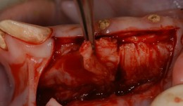
Vertical Ridge Augmentation
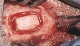
Vertical augmentation is essential for restoring bone defects in patients with destructive gum disease, before rigid fixation of implants or grafts. However, in practice it is associated with high complication rates and limited success. Thus it is important to refine the techniques used by dental surgeons through preclinical research. This chapter looks at various animal studies, and focuses on one specific validated, reproducible and reliable protocol. This involves creating a saddle-like, bony, critical-size defect in the mandible of dogs, in which sufficient time is allowed for the defect to become chronic, thus mimicking bone atrophy in humans. Vertical bone gain is assessed following placement of a tissue-engineered block. Surgical and flap management techniques can be tested, as well as different biologics, devices, scaffolds, membranes, implants and screws. Assessment relates to the initial defect, as well as responses over time relating to resorption of graft particles, soft tissue swelling and inflammation and bone destruction and formation. Osseointegration is quantified to indicate success or failure of the intervention. The protocol can also be used to compare analytical methods. The methods have fairly predictable outcomes, and are applicable to patients with traumatic tooth extraction, jaw damage, endodontic infections and failed implants. -
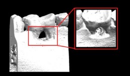
Sinus Floor Augmentation
The maxillary sinuses are located inside the cheekbones, above the upper jaw, from the second premolar area to the wisdom teeth. In some people with tooth loss, the sinuses are too close to the upper jaw for dental implants to be placed; in others, bone may have been reabsorbed because of gum disease. In either case, bone height can be restored using a sinus lift – an increasingly common technique in dental practice. A small hole is made in the bone beneath the gum and the membrane lining the sinus is pushed away from the jaw to create a space into which bone graft material can be packed. Implants can then be placed after the graft has integrated with the natural tissue. Preclinical models are necessary for testing bone grafts from different sources, and different bone substitution materials, biologically active coatings, growth factors, and implant types. The vascularization process can also be investigated. This chapter discusses the merits of various animals for studying the pathology and repair of sinus defects, with a preference for those with similar sinus structures, bone loading characteristics and remodeling processes to humans. Osseointegration of graft materials can take many months, so the protocols here provide a framework of suitable timings and sequences of surgical procedures, including the placing of implants at same time as augmentation. Suitable endpoints are defined too to assist investigators in planning and reporting on reproducible, relevant outcomes. -

Examiner Standardization And Calibration
based on the book chapter by Hatice Hasturk and Mary Ann Cugini Summary The emphasis of this chapter is on examiner--therapist alignment and standardization when conducting controlled clinical trials, especially those with multicenter settings. It begins with definitions of various key research team members and follows by giving specific advice on assessing regeneration by periodontal and bone probing, with reference to parameters relating to probing depth, the cemento--enamel junction, gingival margins, keratinized gingiva, tooth mobility, clinical attachment and alveolar bone gain. The authors explain the relevance of bleeding, plaque and gingival health indices, and the stress the importance of selecting suitable reference points and scoring scales. Detail is provided on the techniques of mucogingival regeneration, alveolar ridge augmentation and implant insertion, and factors such as wound healing. There are protocols for alignment and agreement sessions, and recommendations for conducting regular realignment sessions, with tailored training and calibration, in order to ensure consistency in skills and performance of the team, in data capture and interpretation, and in use of instruments and scoring systems. Practical tips are given on various aspects of the study including set up and timings, examiner--observer agreement and inter-class correlation, with guidance on organizing the research team, assessment plans and statistical analysis. The overall aim of the chapter is to ensure production of high-quality data with minimal uncertainty. Open full-text PDF (1.4 MB) -

Patient-Reported Outcome Measures
based on the book chapter by Colman McGrath Summary There has been a surge of interest in patient-reported outcome measures (PROMs) in all areas of clinical research. Assessing the patients own perceptions of their health, quality of life, functional ability and experience of pain provides very valuable information on the success of an intervention. This chapter describes the development of some commonly used instruments, and summarizes their limitations and suitability for different studies in implant surgery and tissue regeneration. It advises investigators on the selection of different types generic, condition-specific, dimension-specific and utility measures. The authors explain why general (global) instruments like the popular Short Form SF-36 questionnaire have limited sensitivity for oral outcomes, suggesting several condition-specific tools that yield far more specific data and are quick to complete, making them suitable for studies in busy clinic settings and large numbers of patients. The authors describe the role of health utility indexes, which allow patients to rank the importance of items affecting their quality of life, or permit costbenefit analysis of an intervention. The instruments are discussed in the context of their appropriateness for a particular study, their acceptability to patients, validity, reliability and reproducibility, and their responsiveness to change. In terms of interpretation, emphasis is placed on the challenge of identifying the minimally clinically important difference (MCID). Open full-text PDF (0.9 MB)