


-
- English
- Deutsch
- Spanish
-
- General Diagnostics
- Treatment
- Tools/Materials
- Basic/General Knowledge
- Research
- Events
- Organisations
-
-
-
-
-
-

-
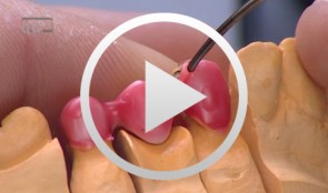

Zirconia Ceramic Restorations Vennered Using the Overpress Technique, part 2
Bußmeier, UweMaterials Checklist: Zeno Tec System Wieland Software: Dental Designer by 3shape Milling unit: Wieland 4030 Zirconia: ZENO® Zr; zirconia staining: Zircolor Investment material: PressX Zr Investment Press: IMAGINE Press with disposable plungers Ingots: PressXZR SHO-3; veneering ceramic: ZIROX Glaze: PressXZR Body Stain A3 / Glaze -
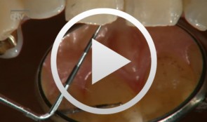
Regenerative Treatment of Class II Mandibular Furcation Defects
Heinz, BerndProcedure Case description: -Class II furcation defect at teeth 46 and 47 and gingival recessions at teeth 43 and 44 - Root planing using PerioSet - Incision technique - Cleaning furcation defect at tooth 46 - Pref Gel application, rinsing and Emdogain application - Insertion of Bio-Oss into the furcation space with an amalgam plugger after hydration - Condensation of the bone replacement material and application of an absorbable membrane (Bio-Gide) - Atraumatic suture closure using 6/0 Seralene Contents: This video demonstration shows the simultaneous treatment of recessions at teeth 43 and 44 and of class II furcation defects at teeth 46 and 47. After a brief case description, root planning is done using PerioSet. Next, an incision is made and the furcation defects are very carefully cleaned using hand instruments and ultrasonic scalers (Soniflex). The cleaned root surfaces and furcation defects are conditioned with Pref Gel (Straumann) for two minutes. The objective of conditioning is to remove the smear layer, to open the dentine tubules, and to enable surface demineralization. Moreover, this measure serves to optimize the contact between Emdogain and the root surface. After two minutes, the EDTA suspension is removed using physiological saline solution or water spray. Immediately afterwards, Emdogain is applied to the blood and saliva-free root surface. This procedure was also used to treat the furcation defect at tooth 47. Regenerative treatment of tooth 46 was performed since that tooth had a very extensive furcation defect. The defect was filled with Bio-Oss, which was applied using an amalgam plugger. Absorbable Bio-Gide was used for coverage of the furcation entrance. Finally, the wound was closed using loop sutures and single interrupted sutures. -
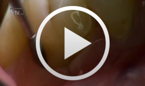
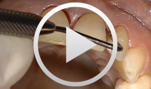
Incisal Edge Restoration and Repair
Frankenberger, RolandProcedure: Incisal Edge Restoration and Repair - Description of defect and problems related to its repair - Airborne particle abrasion and adhesive pretreatment - Biomimetic layering with enamel and dentine mass - Refining, production of halo effect, polishing Contents: The cervical fracture surface lies in the composite region of an underlying Black III cavity. In the past, this completely intact filling probably would have also been removed because of the fracture. Today, it is possible to treat the problem as a repair and leave the neighboring composite restoration in place while maintaining strict adherence to minimally invasive procedure. Even with the help of magnifying glasses, complete removal of the filling probably would have led to enlargement of the defect since the filling had been in place for 10 years. We therefore opted to pretreat the target area near the composite by means of intraoral airborne particle abrasion (Micro-Etcher system using 27 µm Al2O3 powder) before applying phosphoric acid to the enamel and dentine for conventional conditioning. This was followed by the bonding procedure, comprising the application of dentine adhesive to enamel, dentine and the aged composite material. This technique has already been described in the recent literature (Frankenberger et al. Am J Dentistry, 2003). The reconstruction was performed using an enamel and dental mass of esthetic composite material with the help of a silicone key positioned palatinally. This makes it possible to achieve a biomimetic and natural restoration, including a halo effect. Last photograph: Next recall. -
Flap designs for Interdental Tissue Preservation in Periodontal Therapy
Salvi, Giovanni E.Procedure: - Introduction: History -Taking, Examination, Diagnosis, Etiology, Prognosis for Individual Teeth - Four - Phase Treatment Sequence - Modified Papilla Preservation Technique(MPPT) - Simplified Papilla Preservation Technique (SPPT) - Findings 6 months after Surgery Synopsis The Modified and Simplified Papilla Preservation Techniques for conservation of interproximal papillary tissue were designed to provide access to deep and narrow bony defects to enable regenerative periodontal treatment. The Modified Papilla Preservation Technique (MPPT) was designed to ensure tension-free primary closure via barrier membranes in patients with small interdental spaces. The Simplified Papilla Preservation Technique(SPPT) is used to gain access to narrow interdental spaces ( < 2mm) and to deep defects in the lateral tooth region. Apart from preserving primary wound closure in the interdental space, the two techniques also serve to keep the membrane from collapsing into the bony defect. Both MPPT and SPPT employ special suture techniques to ensure tension-free primary closure of the interdental space. This video clip also serves to demonstrate that the two techniques of interproximal tissue preservation can also be used for periodontal interventions without regenerative measures. -
Composite Restoration in Anterior Teeth
Hugo, BurkardProcedure: - Introduction with control of the Gap Width and Colour Selection - Preparation of the Dental Surface (application of the Matrix) - Gap Closure, Final Polish Contents The present case shows the gap closure at tooth 22 with a direct adhesive technique. The gap closure is done here after the orthodontic treatment of a central diastema. A special matrix technique is used to allow a perfect design of the aproximal surfaces and the creation of the aproximal contact points. During the colour selection the dentin and enamel colours are chosen very carefully, to allow a good esthetic result and different composites are used to achieve a natural aspect of the tooth. -
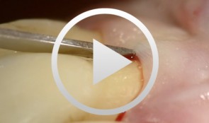
Regenerative Procedures for Optimized Esthetics at Tooth 11
Schlee, MarkusContents: - Exploration - Incision and Flap Mobilization - Palatal Flap Preservation with Interdental Tissue Preservation - Detoxification and Concrement Removal at 11 - Harvesting of Autogenous Bone Chips from the Spina Nasalis - Conditioning of the Root Surface with EDTA-Gel - Application of Emdogain and Filling of the Bone Defect - Wound Closure Synopsis After Finishing the Initial Treatment for Aggressive Periodontitis, Regenerative Treatment of a Tunnel-Shaped Pocket at Tooth 11 was attempted. Rotation and Crowding of the Buccally Inclined Tooth represented a favorable Etiological Factor. The patient did not wish to receive Orthodontic Treatment to eliminate this Causal Factor after Completion of Primary Treatment. Treatment was therefore limited to the Surgical Regeneration Attempt. The Interdental Space was larger than 3 mm and the Bone Pocket was a mostly Three-Walled Structure, so the Chances of Success were considered to be good. Exploration was first performed to identify the Course of the Defect Margins. Exact knowledge of the Bone Anatomy in all three Planes is essential to successful Incision Planning. A Tunnel-Shaped Defect delimited by Bone in the Region of Tooth 11 with good chances of Periodontal Regeneration was found. A major Challenge of this Procedure is the need to keep the Defect completely covered with Soft Tissue throughout the Healing Process. Cortellini's Papilla Preservation Technique was used for this Purpose. After Incision and Flap Mobilization, it became evident that the Defect only had two Walls in the Coronal Region and that Bone was lacking in the Buccal Region. According to the current Data on Periodontal Regeneration, the Attachment Gain achieved using an Enamel Matrix Protein (Emdogain®) alone can be just as good as that achieved using Emdogain and Bone Graft Material combined. Still, we elected to use a Combination Technique in the Present Case because it provides better Papillary Support. The Graft Material consisted of Autogenous Bone Chips from the Spina Nasalis, which can easily be harvested by Means of the Piezo Technique After gentle Detoxification, the Root Surface was treated with Emdogain. The Defect was then filled with Autogenous Bone Chips and closed by Microsurgical Suture Techniques. Six months after Surgery, Partial Regeneration of the Papilla can be seen. -
Regenerative Measures for Osseous Defect Repair and Optimal Esthetics
Sculean, AntonProcedure: Theoretical Part: - Adult male with a deep and broad intraosseous bone defect located on tooth #13 - The indication for modified papilla preservation in the scope of regenerative therapy was established based on the width of the diastema - Regenerative periodontal therapy with Emdogain and a Bio-Oss® cancellous bone graft - Emdogain is applied to the root surface to stimulate regeneration of periodontal structures - To prevent graft collapse and to minimize the risk of development of too large a recession in this esthetically important region, the defect was filled with Bio-Oss® cancellous bone material Practical Part: - The papilla preservation technique was performed using microsurgical instruments - The root surface area was conditioned with 24% EDTA for ca. 2 minutes - Emdogain was applied to the root surface - The defect was filled with the Emdogain/Bio-Oss® mixture - The wound was closed with two mattress sutures one horizontal mattress suture to secure the graft in place, and a second modified vertical mattress suture to tightly close the papilla - A 5-0 suture was used for the horizontal mattress suture, and a 6-0 monofilament was used for the vertical mattress suture - Postoperative care entailed rinsing the wound twice daily for 4 weeks with 0.2% chlorhexidine and ibuprofen analgesia on the first few days after surgery Contents: The patient's jaw displayed a generalized loss of clinical attachment and alveolar bone. His general history was unremarkable; the patient was a non-smoker. Microbiological tests showed large numbers of Actinobacillus actinomycetemcomitans and Porphyromonas gingivalis. The diagnosis was "generalized aggressive periodontitis". After four months of initial therapy consisting of antibiotic combination therapy (amoxicillin + metronidazole), intraoral radiographs showed a deep and wide intraosseous bone defect located mesial and palatal to tooth #13. To preserve this strategically important tooth we opted to perform regenerative therapy with Emdogain and Bio-Oss cancellous bone material. Ten months after regenerative periodontal therapy, the probing depth had decreased by 7 mm, and 5-6 mm of clinical attachment had been gained. At this time, the probing depth was 2-3 mm and intraoral radiographs showed near-complete filling of the osseous defect. -
Intraorally Fabricated, Glass Fiber Reinforced Composite Bridge for Replacement of Individual Anterior Teeth - The entire case
Hugo, BurkardProcedure: - Introduction and establishment of indication for the treatment - Bridge construction with glass fiber reinforcement - Presentation of the patient with congenital absence of teeth # 12 and 22 - Clinical procedure for fabrication of a bridge for tooth #22 - Discussion of the technique Contents: Here, we describe a technique for direct application of composite bridges with adhesive bonding. The procedure is designed to provide a replacement for individual anterior teeth, especially in younger individuals who have lost a tooth due to trauma or who have congenitally missing teeth. Bridge manufacture is completely intraoral. By using a one or two-wing abutment design, a framework of parallel, pre-impregnated glass fibers is adhesively bonded, and the midsection is freely built up from composite by a special procedure. This systematic approach permits reasonably priced manufacture of direct tooth replacements with predictably good esthetic results. Maximal conservation of substance and reversibility, which is achieved by dispensing with prepping measures, means that the procedure does not limit the possibilities for future restorations (e.g., implants). -
Microsurgical Removal of a Foreign Body from the Mandibular Canal
Schultze-Mosgau, StefanOverview: - Access and incision: Creation of a vestibular pedicled mucoperiosteal flap via a gingival margin incision while preserving the papilla - Removal of vestibular bone in the region of tooth 46 using a microsurgical instrument - Exposure of the neurovascular bundle - Removal of the foreign body - Re-adaptation of the mucoperiosteal flap - Wound closure with atraumatic suture material Contents: Female patient with an indication for microsurgical foreign body removal (removal of a fractured root canal instrument from a previous endodontic treatment of tooth 46) using a surgical microscope. The foreign body extends from the apex into the mandibular canal. -
Surgical Treatment of Periodontitis Using a Minimally Invasive Approach
Beck, FrankThis case is an excellent demonstration of the use of the minimally invasive access flap technique for treatment of (chronic) periodontitis in an esthetically critical zone. The access flap was used in conjunction with enamel matrix proteins for regenerative therapy.,