


-
- English
- Deutsch
- Spanish
-
- General Diagnostics
- Treatment
- Tools/Materials
- Basic/General Knowledge
- Research
- Events
- Organisations
-
-
-
-
-
-

-
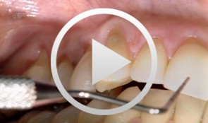

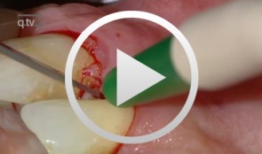
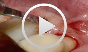
Recession coverage with coronally positioned flap
Zucchelli, GiovanniOutline: - Aesthetic treatment of multiple gingival recessions List of materials Microblade BUSM-6700 (Stoma) and 15C Swan Morton, Periosteum elevator Free, De Wijs, Molt (Stoma), Curettes minifive 11-12, 7-8 (LM), Pinzette stright 17cm 0,6 (stoma), Neddle holder micro Barraquer straight 18cm extra fine (stoma), Micro-scissor Gomel, curve, 16cm extra fine (stoma), Suture Vicryl 6-0 (V384) -
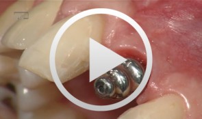
Implant-supported crown at site 21
Weigl, Paul / Trimpou, Georgia -
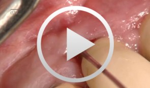
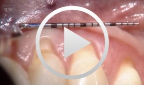
Laser decontamination at sites 11 and 12
Schwarz, FrankOutline: - Presentation of clinical findings - Explanations Er:YAG laser/fibre tip/power settings - Treatment - Postoperative management List of materials: Er:YAG laser device (KEY 3®, KaVo, Biberach, Germany) Laser handpiece (2061, KaVo, Biberach, Germany) Cone-shaped glass fibre tip Safety goggles Periodontal probe (PCP12, Hu-Friedy Co., Chicago, Illinois, USA) -
Recession coverage using human dermal tissue at site 22-26
Schlee, Markus -
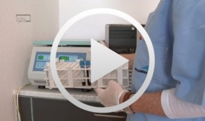
The use of autologous PRGF in periodontal plastic surgery
Marggraf, ErwinContents: - Blood sampling - Platelet separation activation - Introduction of bone replacement material - Access flap and curettage - Introduction of PRGF and bone replacement material - Plastic suture Materials Checklist: All materials required for producing PRGF (BTI Germany) Bone replacement materials Geistlich Biomaterials Surgical instruments, Aesculap Suture materials, Ethicon -
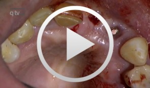
Periodontal regeneration at teeth 21 and 23 using EMD and cortical bone chips
Topoll, Heinz H.Contents: - Incisions using a microsurgical scalpel - Reflecting a buccal flap - Preparing papillary flaps using a microsurgical scalpel - Lifting off of the papillary flaps using a papillary elevator - Removing the granulation tissue using an ultrasound scaler - Cleaning the dental roots using manual instruments - Trying to dental root - Applying Emdogain - Mixing Bio-Oss and Emdogain - Introducing the Bio-Oss into both bone defects - Microsurgical suturing Materials Checklist: Cheek retractor Microsurgical scalpel blade holder Microsurgical scalpel blade Soniflex tips Bone rest Castroviejo microsurgical needle holder Suturing scissors Dental tweezers Microsurgical tweezers Monofilament suturing material 6/0 Seralene Pref gel Emdogain Bio-Oss -
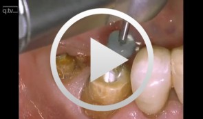
Core & Post System Post Cementation and Core Build Up
Nölken, RobertMaterials Checklist: Dentsply Core&Post system kit. -
Ridge augmentation in the periodontally involved dentition
Windisch, PéterContents: - Periodontal regeneration and alveolar -ridge augmentation using a connectivetissue graft - Implant insertion and augmentation - Implant re-entry and prosthetics Materials Checklist Emdogain, Bio-Oss, BioGide, Block fixating screw for autologous bone cylinder, 4/0 and 5/0 sutures, Resolut membrane Titanium pins, Autologous bone chips, 2 Replace Groovy Tapered 4, 3x13 mm implants -
Flap surgery using a modified papilla preservation technique
Salvi, Giovanni E.Outline - Introduction: Patient history, clinical findings, diagnoses, etiology, prognosis of individual teeth - Treatment planning for phases - Modified papilla preservation technique (MPPT) - Simplified papilla preservation technique (SPPT) -Clinical situation 6 months postoperatively -
Operative Therapiekonzepte zur Entfernung retinierter unterer Weisheitszähne
Schultze-Mosgau, Stefan / Neukam, Friedrich Wilhelm / Basting, GerdGliederung: - Schematische und röntgenologische Demonstration unterschiedlicher Retentionsformen unterer, retinierter Weisheitszähne, Darstellung der Indikation zur Entfernung - Erstellung der Behandlungsunterlagen, Aufklärungsgespräche mit Darstellung der Komplikationen - Demonstration des operativen Vorgehens: Lokalanästhesie, Schnittführung, Schutz des N. lingualis, Osteotomie, Nahttechnik. Die Entfernung unterer, retinierter Weisheitszähne gehört zu den häufigsten dento-alveolär-chirurgischen Eingriffen. Durch die enge anatomische Lagebeziehung zu den benachbarten Zähnen und dem N. alveolaris inferior besteht bei der operativen Entfernung die Gefahr einer Schädigung der umgebenden Strukturen. Die Kenntnis der verschiedenen Retentionsformen und eine geeignete, atraumatische Operationstechnik ist für eine komplikationslose Entfernung von Bedeutung. Nach der Leitungsanästhesie des N. alveolaris inferior und des N. buccalis wird die Schnittführung so gewählt, dass ein vestibulär gestielter Mukoperiostlappen gehoben werden kann. Nach dem lingualen, subperiostalen Einführen eines Raspatoriums zum Schutz des N. lingualis wird durch die bukkale Osteomie mit kugelförmigen Hartmetallfräsen der Weisheitszahn bis zu seiner größten Zirkumferenz freigelegt und durch vorsichtige Luxationsbewegungen entfernt.