


-
- English
- Deutsch
- Spanish
-
- General Diagnostics
- Treatment
- Tools/Materials
- Basic/General Knowledge
- Research
- Events
- Organisations
-
-
-
-
-
-

-
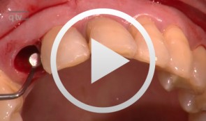

Immediate Function with NoblePerfect Implants
Nölken, RobertProcedure: - Extraction of upper front teeth not suitable for preservation - Immediate loading of four NobelPerfectTM implants - Intraoperative registration of implant position - Flap-free Vestibular Bone Augmentation - Connective Tissue Graft for Periodontal Regeneration - Immediate Loading of temporary implants on the day of surgery - Definitive restoration 6 months later Materials: NobelPerfect Groovy Implants RP and NP; NobelPerfect Temporary Abutments RP and NP; NobelPerfect Implant Replicas RP and WP; Ribbond, Bondable Reinforcement Ribbon; Ethilon 5-0 FS3 Suture Material; Astra Bonetrap Bone Collector. -
-

Ballon Lift Control - A life report
Heuckmann, Karl-Heinz -
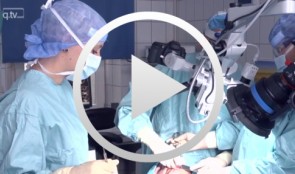
Microsurgical lateral sinus floor elevation (LSFE)
Nölken, RobertOutline: - Incision - Flap mobilization - Lateral sinus fenestration - Elevation of the Schneiderian membrane - Implant bed preparation - Bone chip harvesting at the mandibular angle - Filling of sinus lift lumen with autologous bone chips - Implant insertion - Covering the lateral sinus cavity with collagen membrane - Wound closure List of materials - Zeiss Pro Dent microscope with beam splitter and Panasonic 3 CCD camera - Scalpel holder (Ustomed) with Swann-Morton blades 15C and 12D - Narrow rasp (Hu-Friedy) - Micro-vacuum (Luer Lock Suction Tip, American Dental Systems) - Disposable vacuum tube set (Bexamed) - Disposable draping, Lindau (Aescologic) - Piezosurgery with diamond ball (Mectron) - Microforceps (Hu-Friedy) - Excavator (Martin) - Periodontometer, 1-mm gradation (Hu-Friedy) - OsseoSpeed implant set, Dentsply Implants: Marking drill; Twist drill, 2 mm; Depth gauge; Pilot drill, 2/3.2 mm; Twist drill, 3.2 mm; Tapered drill, 3.2/5 mm; OsseoSpeed TX implant, 5.0 × 11 mm; Closure screw, 4.5/5 mm - Columbia curette (Ustomed) - Micross scraper (Meta) - Needle holder (Ustomed) - Langenbeck wound retractor (Ustomed) - Kelly scissors (Ustomed) - Buchanan endodontic hand plugger (American Dental Systems) - Resorbable collagen membrane (Resodont, Resorba) - Ethilon 5-0 FS-3 (Ethicon) - Prolene 6-0 DA-2 (Ethicon) -
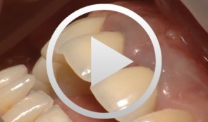
Regenerative Measures for Osseous Defect Repair and Optimal Esthetics
Sculean, AntonProcedure: Theoretical Part: - Adult male with a deep and broad intraosseous bone defect located on tooth #13 - The indication for modified papilla preservation in the scope of regenerative therapy was established based on the width of the diastema - Regenerative periodontal therapy with Emdogain and a Bio-Oss® cancellous bone graft - Emdogain is applied to the root surface to stimulate regeneration of periodontal structures - To prevent graft collapse and to minimize the risk of development of too large a recession in this esthetically important region, the defect was filled with Bio-Oss® cancellous bone material Practical Part: - The papilla preservation technique was performed using microsurgical instruments - The root surface area was conditioned with 24% EDTA for ca. 2 minutes - Emdogain was applied to the root surface - The defect was filled with the Emdogain/Bio-Oss® mixture - The wound was closed with two mattress sutures one horizontal mattress suture to secure the graft in place, and a second modified vertical mattress suture to tightly close the papilla - A 5-0 suture was used for the horizontal mattress suture, and a 6-0 monofilament was used for the vertical mattress suture - Postoperative care entailed rinsing the wound twice daily for 4 weeks with 0.2% chlorhexidine and ibuprofen analgesia on the first few days after surgery Contents: The patient's jaw displayed a generalized loss of clinical attachment and alveolar bone. His general history was unremarkable; the patient was a non-smoker. Microbiological tests showed large numbers of Actinobacillus actinomycetemcomitans and Porphyromonas gingivalis. The diagnosis was "generalized aggressive periodontitis". After four months of initial therapy consisting of antibiotic combination therapy (amoxicillin + metronidazole), intraoral radiographs showed a deep and wide intraosseous bone defect located mesial and palatal to tooth #13. To preserve this strategically important tooth we opted to perform regenerative therapy with Emdogain and Bio-Oss cancellous bone material. Ten months after regenerative periodontal therapy, the probing depth had decreased by 7 mm, and 5-6 mm of clinical attachment had been gained. At this time, the probing depth was 2-3 mm and intraoral radiographs showed near-complete filling of the osseous defect. -
-
Cell-to-Cell Communication: Periodontal Regeneration
Stadlinger, Bernd / Terheyden, HendrikUnlike bone, which undergoes remodeling by resorption followed by bone apposition, teeth are not subject to physiological remodeling. They are remarkably resistant to physiological remodeling processes. The reason for this is found in the periodontium and cementum. The mechanisms that take effect in this area and the effectiveness of the periodontal system - consisting of gingiva, alveolar bone, periodontium and cementum - after injuries will be visualized in the new scientific 3D film, "Periodontal Regeneration".The visualization of these complex processes is of great interest for academic teaching as well as for the clinician and general practitioner as the cellular interactions are presented in the context of four phases. For the first time scanning electron microscopic images of real cells will be visualized allowing the comparison of their characteristics with computer animated simulations. DVD 1: Expert Version approx. 14 minutes DVD 2: Public Version approx. 13 minutes Outline: - Cementum formation - Orthodontic tooth movement - Trauma and periodontitis - Periodontal regeneration -

Periodontal Preserve Therapy (Examples)
Clotten, StefanContent: - Periodontal maintenance therapy for teeth 34 and 35, including the regeneration of a bone defect using bone replacement material, collagen membrane and sutures. - Curettage for treatment of periodontal pockets. - Treatment of gingival pressure sores caused by tight-fitting orthodontic apparatus. - Incision of buccal attachment to relieve gingival pressure for elimination of gingival recession. -
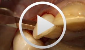
Treatment of a palatal class II furcation
Marggraf, ErwinOutline: - Reflecting a flap - Cleaning. - Fraction 3 - Fraction 2 - Fraction 1 - Wound closure List of materials All materials required for producing PRGF (BTI Germany) Bone replacement materials Geistlich Biomaterials Surgical instruments, Aesculap Suture materials, Ethicon -
The use of autologous PRGF in periodontal plastic surgery
Marggraf, ErwinContents: - Blood sampling - Platelet separation activation - Introduction of bone replacement material - Access flap and curettage - Introduction of PRGF and bone replacement material - Plastic suture Materials Checklist: All materials required for producing PRGF (BTI Germany) Bone replacement materials Geistlich Biomaterials Surgical instruments, Aesculap Suture materials, Ethicon