


-
- English
- Deutsch
- Spanish
-
- General Diagnostics
- Treatment
- Tools/Materials
- Basic/General Knowledge
- Research
- Events
- Organisations
-
-
-
-
-
-

-
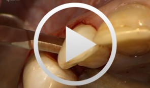

Regenerative Measures for Osseous Defect Repair and Optimal Esthetics
Sculean, AntonProcedure: Theoretical Part: - Adult male with a deep and broad intraosseous bone defect located on tooth #13 - The indication for modified papilla preservation in the scope of regenerative therapy was established based on the width of the diastema - Regenerative periodontal therapy with Emdogain and a Bio-Oss® cancellous bone graft - Emdogain is applied to the root surface to stimulate regeneration of periodontal structures - To prevent graft collapse and to minimize the risk of development of too large a recession in this esthetically important region, the defect was filled with Bio-Oss® cancellous bone material Practical Part: - The papilla preservation technique was performed using microsurgical instruments - The root surface area was conditioned with 24% EDTA for ca. 2 minutes - Emdogain was applied to the root surface - The defect was filled with the Emdogain/Bio-Oss® mixture - The wound was closed with two mattress sutures one horizontal mattress suture to secure the graft in place, and a second modified vertical mattress suture to tightly close the papilla - A 5-0 suture was used for the horizontal mattress suture, and a 6-0 monofilament was used for the vertical mattress suture - Postoperative care entailed rinsing the wound twice daily for 4 weeks with 0.2% chlorhexidine and ibuprofen analgesia on the first few days after surgery Contents: The patient's jaw displayed a generalized loss of clinical attachment and alveolar bone. His general history was unremarkable; the patient was a non-smoker. Microbiological tests showed large numbers of Actinobacillus actinomycetemcomitans and Porphyromonas gingivalis. The diagnosis was "generalized aggressive periodontitis". After four months of initial therapy consisting of antibiotic combination therapy (amoxicillin + metronidazole), intraoral radiographs showed a deep and wide intraosseous bone defect located mesial and palatal to tooth #13. To preserve this strategically important tooth we opted to perform regenerative therapy with Emdogain and Bio-Oss cancellous bone material. Ten months after regenerative periodontal therapy, the probing depth had decreased by 7 mm, and 5-6 mm of clinical attachment had been gained. At this time, the probing depth was 2-3 mm and intraoral radiographs showed near-complete filling of the osseous defect. -
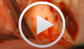
Microsurgical Removal of a Foreign Body from the Mandibular Canal
Schultze-Mosgau, StefanOverview: - Access and incision: Creation of a vestibular pedicled mucoperiosteal flap via a gingival margin incision while preserving the papilla - Removal of vestibular bone in the region of tooth 46 using a microsurgical instrument - Exposure of the neurovascular bundle - Removal of the foreign body - Re-adaptation of the mucoperiosteal flap - Wound closure with atraumatic suture material Contents: Female patient with an indication for microsurgical foreign body removal (removal of a fractured root canal instrument from a previous endodontic treatment of tooth 46) using a surgical microscope. The foreign body extends from the apex into the mandibular canal. -
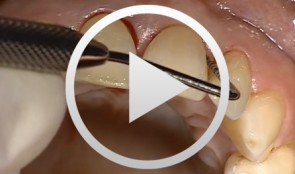
Surgical Treatment of Periodontitis Using a Minimally Invasive Approach
Beck, FrankThis case is an excellent demonstration of the use of the minimally invasive access flap technique for treatment of (chronic) periodontitis in an esthetically critical zone. The access flap was used in conjunction with enamel matrix proteins for regenerative therapy., -
-
SOS - An innovative method for the implantological rehabilitation of the edentulous mandible
Sliwowski, Christoph T. -
Implant placement in the lower posterior area with immediate provisionalization
Hürzeler, Markus B. -
-
Microsurgical lateral sinus floor elevation (LSFE)
Nölken, RobertOutline: - Incision - Flap mobilization - Lateral sinus fenestration - Elevation of the Schneiderian membrane - Implant bed preparation - Bone chip harvesting at the mandibular angle - Filling of sinus lift lumen with autologous bone chips - Implant insertion - Covering the lateral sinus cavity with collagen membrane - Wound closure List of materials - Zeiss Pro Dent microscope with beam splitter and Panasonic 3 CCD camera - Scalpel holder (Ustomed) with Swann-Morton blades 15C and 12D - Narrow rasp (Hu-Friedy) - Micro-vacuum (Luer Lock Suction Tip, American Dental Systems) - Disposable vacuum tube set (Bexamed) - Disposable draping, Lindau (Aescologic) - Piezosurgery with diamond ball (Mectron) - Microforceps (Hu-Friedy) - Excavator (Martin) - Periodontometer, 1-mm gradation (Hu-Friedy) - OsseoSpeed implant set, Dentsply Implants: Marking drill; Twist drill, 2 mm; Depth gauge; Pilot drill, 2/3.2 mm; Twist drill, 3.2 mm; Tapered drill, 3.2/5 mm; OsseoSpeed TX implant, 5.0 × 11 mm; Closure screw, 4.5/5 mm - Columbia curette (Ustomed) - Micross scraper (Meta) - Needle holder (Ustomed) - Langenbeck wound retractor (Ustomed) - Kelly scissors (Ustomed) - Buchanan endodontic hand plugger (American Dental Systems) - Resorbable collagen membrane (Resodont, Resorba) - Ethilon 5-0 FS-3 (Ethicon) - Prolene 6-0 DA-2 (Ethicon) -
Sinus Floor Augmentation with Autogenous Chin Bone Grafts
Schultze-Mosgau, Stefan / Neukam, Friedrich Wilhelm / Basting, GerdContent: In the maxillary incisor region, a sinus floor augmentation to enlarge the vertical bone supply may be indicated for a vertically reduced local bone height of less than 5 to 7 mm before procedures to rehabilitate masticatory function with an implant-bearing tooth replacement. For a single-sided deposit osteoplasty, the quantity of autogenous bone from the chin region is usually sufficient. The operative procedure of a single-sided lateral sinus floor augmentation is demonstrated with particulate spongious bone and alternatively with an autogenous block graft. The video also shows the operative method for a crestal sinus floor augmentation with the aid of the endoscopically controlled condensation technique. The advantages and disadvantages of the individual procedures are highlighted. In addition, the technique for harvesting chin bone transplants in different case examples is shown. Outline: - Operative technique for lateral sinus floor augmentation with autogenous particulate spongious bone - Operative technique for lateral sinus floor augmentation with autogenous block grafts - Crestal, endoscopically controlled sinus floor augmentation with condensation technique - Techniques for harvesting chin bone grafts - Range of indication for sinus floor augmentation - Lateral sinus floor augmentation - Operative technique of crestal, endoscopically controlled sinus floor augmentation - Operative technique of autogenous chin bone removal -
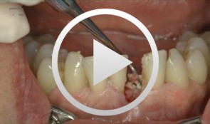
Defect Prevention following Extraction of a Maxillary Central Incisor
Zuhr, OttoContents: - Minimally invasive, atraumatic extraction of an anterior tooth - Buccal soft tissue augmentation using a modified tunneling technique - Socket preservation technique for conservation of the extraction socket - Provisional restoration and closure using modified suspension sutures Materials Checklist: Tunneling Knife® (Dr. Zuhr), No. 1 / No. 2 Keydent Microblade SR Geistlich Bio-Oss® Spongiosa, particle size 0.25 - 1 mm Geistlich Bio-Gide® membrane, 25 x 25 mm Seralene Blue 7/0 DS-15, 0.5 m sutures CV-5 Gore-Tex sutures