


-
- English
- Deutsch
- Spanish
-
- General Diagnostics
- Treatment
- Tools/Materials
- Basic/General Knowledge
- Research
- Events
- Organisations
-
-
-
-
-
-

-

-
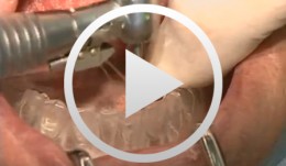
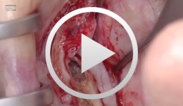
Microsurgical lateral sinus floor elevation (LSFE)
Nölken, RobertOutline: - Incision - Flap mobilization - Lateral sinus fenestration - Elevation of the Schneiderian membrane - Implant bed preparation - Bone chip harvesting at the mandibular angle - Filling of sinus lift lumen with autologous bone chips - Implant insertion - Covering the lateral sinus cavity with collagen membrane - Wound closure List of materials - Zeiss Pro Dent microscope with beam splitter and Panasonic 3 CCD camera - Scalpel holder (Ustomed) with Swann-Morton blades 15C and 12D - Narrow rasp (Hu-Friedy) - Micro-vacuum (Luer Lock Suction Tip, American Dental Systems) - Disposable vacuum tube set (Bexamed) - Disposable draping, Lindau (Aescologic) - Piezosurgery with diamond ball (Mectron) - Microforceps (Hu-Friedy) - Excavator (Martin) - Periodontometer, 1-mm gradation (Hu-Friedy) - OsseoSpeed implant set, Dentsply Implants: Marking drill; Twist drill, 2 mm; Depth gauge; Pilot drill, 2/3.2 mm; Twist drill, 3.2 mm; Tapered drill, 3.2/5 mm; OsseoSpeed TX implant, 5.0 × 11 mm; Closure screw, 4.5/5 mm - Columbia curette (Ustomed) - Micross scraper (Meta) - Needle holder (Ustomed) - Langenbeck wound retractor (Ustomed) - Kelly scissors (Ustomed) - Buchanan endodontic hand plugger (American Dental Systems) - Resorbable collagen membrane (Resodont, Resorba) - Ethilon 5-0 FS-3 (Ethicon) - Prolene 6-0 DA-2 (Ethicon) -
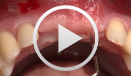
Defect Prevention following Extraction of a Maxillary Central Incisor
Zuhr, OttoContents: - Minimally invasive, atraumatic extraction of an anterior tooth - Buccal soft tissue augmentation using a modified tunneling technique - Socket preservation technique for conservation of the extraction socket - Provisional restoration and closure using modified suspension sutures Materials Checklist: Tunneling Knife® (Dr. Zuhr), No. 1 / No. 2 Keydent Microblade SR Geistlich Bio-Oss® Spongiosa, particle size 0.25 - 1 mm Geistlich Bio-Gide® membrane, 25 x 25 mm Seralene Blue 7/0 DS-15, 0.5 m sutures CV-5 Gore-Tex sutures -
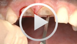
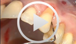
Periodontal regeneration at teeth 21 and 23 using EMD and cortical bone chips
Topoll, Heinz H.Contents: - Incisions using a microsurgical scalpel - Reflecting a buccal flap - Preparing papillary flaps using a microsurgical scalpel - Lifting off of the papillary flaps using a papillary elevator - Removing the granulation tissue using an ultrasound scaler - Cleaning the dental roots using manual instruments - Trying to dental root - Applying Emdogain - Mixing Bio-Oss and Emdogain - Introducing the Bio-Oss into both bone defects - Microsurgical suturing Materials Checklist: Cheek retractor Microsurgical scalpel blade holder Microsurgical scalpel blade Soniflex tips Bone rest Castroviejo microsurgical needle holder Suturing scissors Dental tweezers Microsurgical tweezers Monofilament suturing material 6/0 Seralene Pref gel Emdogain Bio-Oss -

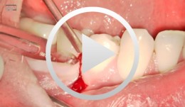
The use of autologous PRGF in periodontal plastic surgery
Marggraf, ErwinContents: - Blood sampling - Platelet separation activation - Introduction of bone replacement material - Access flap and curettage - Introduction of PRGF and bone replacement material - Plastic suture Materials Checklist: All materials required for producing PRGF (BTI Germany) Bone replacement materials Geistlich Biomaterials Surgical instruments, Aesculap Suture materials, Ethicon -
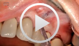
Implants for the Anterior Maxillary Region
Horrichs, Leon G.Contents: - Implant planning and positioning - Preparation - Drilling and thread-cutting - Implant insertion - Wound closure Synopsis: The implant position is determined using a drill guide/x-ray template (regions 34-32-42-44). A Peeso drill is inserted in the drill guide and used to mark the position in the mucoperiosteum. An incision is made, and the mucoperiosteum is displaced. The implant insertion sites are prepared by using a 2 mm twist drill, 2/3 mm pilot drill, 3 mm and 3/5 mm twist drills. This is followed by counter-drilling and thread-cutting. The implants were loaded at 40-50 N. The wound is then closed using GORE-TEX® suture material. -
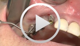
Microsurgical apical resection on a maxillary premolar
Nölken, Robert -
Socket-shield surgery on two central incisors
Hürzeler, Markus B. -
Microsurgical apical resection
Nölken, Robert -
-

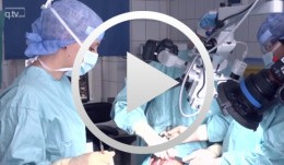
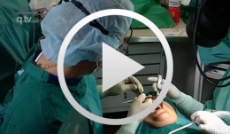
Live surgery Surgical treatment of bone necrosis
Schultze-Mosgau, StefanOutline: - Surgical wound debridement - Sequestrotomy - Preparation of the soft-tissue bed - Plastic, tension-free, saliva-proof wound closure List of materials Basic surgical tool set: - Surgical blade - Preparation scissors - Pair of tweezers - Suture materials -
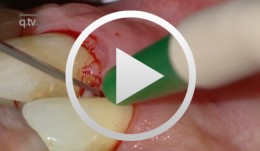
Covering a Recession with a Soft Tissue Transplant
Heinz, Bernd / Jepsen, SörenObjectives: Use of a soft tissue graft for recession coverage at tooth 23 and for gingival augmentation. Content: 1. Incision around tooth 23, intra-sulcular preparation, mobilization of coronal sliding flap, and pre-flap preparation. 2. Root smoothing, reduction of ground cavity with diamond burs from Perioset system. 3. Preparation and harvesting of connective tissue flap from palate, Emdogain application, and wound closure. 4. Placement of interrupted interdental sutures for fixation of connective tissue flap.