


-
- English
- Deutsch
- Spanish
-
- General Diagnostics
- Treatment
- Tools/Materials
- Basic/General Knowledge
- Research
- Events
- Organisations
-
-
-
-
-
-

-

Innovative CAD/CAM treatment approaches for implant-supported fixed restorations
Beuer, Florian / Stimmelmayr, Michael / Schweiger, JosefOutline: - Patient presentation - Preparing the implant bed, implant placement, checking implant positions - Securing the insertion posts to the index for fabrication of the cast - Suturing details - Delivery of the adapted long-term provisional - Fabricating the cast and the gingival mask, transferring the pontic emergence profiles to the gingival mask, mask adaptation - The master cast under the strip scanner with scan bodies on the laboratory analogs - CAD crown design and virtual anatomic shaping - CAM fabrication of a zirconia abutment - Adhesively connecting the zirconia abutment to the titanium base - Reentry, split-thickness flap, vestibuloplasty, connecting the zirconia abutments to the implants - Mucosal graft to restore a soft-tissue defect - Intraoral impression of the abutments - Fabricating the definitive lithium disilicate crowns: virtual crown design; CAM milling, characterization of the crowns - Delivery, final adjustments, presentation of the treatment outcome -
-
12 weeks after a sandwich osteotomy in the posterior mandible: implant insertion
Bormann, Kai-Hendrik -

Errors and Checklists
Franck Renouard -

-

-

-

-

-
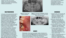
THE ROLE OF MULTIDISCIPLINARY APPROACH IN A CASE OF LANGERHANS CELL HISTIOCYTOSIS WITH INITIAL PERIODONTAL MANIFESTATIONS
Objectives: The present case epitomizes the clinical situation of a single-system Langerhans cell histiocytosis (LCH) mimicking aggressive periodontitis in a patient with no other clinical signs. Although this is a single observation, we highlight the importance of using a multidisciplinary approach in rare conditions like this for optimizing patient management. Methods: A 32-year-old Caucasian woman visited a private dental practice in Brescia, Italy, complaining of sensitivity in her mandibular left first molar. Clinical and radiographic examinations revealed recession, furcation involvement, mobility and severe alveolar bone loss, leading to a diagnosis of localised aggressive periodontitis. Over the next 3 years, the patient received non-surgical periodontal treatment, but failed attend successive follow-up appointments for undisclosed reasons. Her periodontal condition continued to worsen and multiple tooth extractions were carried out due to progressive periodontal destruction with impaired healing and development of ulcerative lesions. Panoramic radiographs were taken, and revealed severe alveolar bone destruction with “floating teeth” appearance, and an osteolytic bone lesion at the left angle of the mandible. With an overall clinical deterioration, and the possibility of an underlying malignant condition, the patient was referred for deep analysis. Results: No abnormalities were detected in laboratory and biochemical tests. Skull and sinus radiography revealed a 5-mm oval radiolucency at the left angle of the mandible. Then a radiograph of the lower leg revealed multiple osteolytic bone lesions of the left tibia. Bone-marrow aspiration and biopsy were performed to determine the nature of these lesions. Histopathology revealed a dense nodular proliferation of CD1a-positive and S100-positive Langerhans cells within bone, on a background of lymphocytic and granulocytic cells, consistent with LCH. An additional biopsy of the intra-oral lesion showed mature, disease-free, compact bone. However, a bone biopsy may be not representative of the entire structure, particularly in cases of intraoral localisation of LCH. Bidirectional Sanger sequencing analysis and pyrosequencing of DNA extracted from bone tissue of the tibia detected the presence of the BRAF-V600E hotspot somatic mutation, confirming a clonal origin of the neoplastic cells. Multidisciplinary investigations showed that the periodontal involvement was a manifestation of an underlying systemic disease (multifocal single-system LCH). The patient was then started on radiotherapy and but improvement of her oral and periodontal condition is yet to be confirmed. Conclusions: In the present case, LCH was unrecognised for several years. The periodontal disease progressed rapidly, leading to loss of most of the dentition, with persistent delays in soft tissues healing after extraction. Close monitoring of the oral signs may have allowed earlier diagnosis of LCH and prevent such rapid deterioration, possibly resulting in a better endpoint. Dentists and periodontists should be aware that rare systemic diseases such as LCH can produce oral manifestations as the first clinical sign. Such patients benefit from a multidisciplinary approach to identify and manage such entities. -
DENTAL IMPLANT SURVIVAL IN HYPODONTIA PATIENTS: A RETROSPECTIVE STUDY
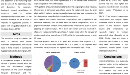
Objectives: Specialists in prosthodontics and restorative dentistry are often challenged by oral rehabilitation of patients with hypodontia. Implant rehabilitation is commonly used to replace developmentally missing teeth, but evidence of implant survival in hypodontia patients is scarce. The aim of this study was to present findings on short- to medium-term clinical outcomes, complications and survival rates of dental implants in patients with hypodontia who were treated in Birmingham, UK. Methods: A retrospective analysis was conducted of the clinical records of patients treated within the Oral and Maxillofacial Surgery Department at Queen Elizabeth Hospital and the Restorative Dentistry Department at the University of Birmingham Dental Hospital. All patients were identified from the hypodontia clinic database. Clinical variables were recorded, collated and compared with reports of adverse events during a follow-up period of up to 6.7 years. Patient data were collected using a pro forma regarding the number and the location of implants, the surgical procedure, and any surgical or restorative complications. All patients on the study had a review appointment within the previous 3 months. The data were analysed to determine the number of patients treated, the type and number of implants placed, the site of placement and type of augmentation used, and to identify any peri-operative and post-operative complications. The minimum, maximum and mean follow-up period was calculated and the number of implants per follow-up year were recorded. Results: There were 67 patients aged between 20 and 57 (average 28 years) who received 304 implants to replace anterior (54.9%) and posterior (45.1%) missing tooth units; 49% of implants were placed in the maxilla and 51% in the mandible; 36.5% were placed in non-grafted sites and 63.5% in grafted sites. Mandibular ramus augmentation was used in 80.3% of cases, iliac crest augmentation in 11.9%, and bovine origin xenografts in 7.8%. Post-surgical complications were experienced by 19.4% of patients; four had fenestration or dehiscence type defects around the implant, the graft failed in two, and seven had transient anaesthesia or paraesthesia in the inferior dental nerve distribution area. There were complications in 3.2% implants after completion of the restorative treatment, 40% of which were soft tissue complications (e.g. gingival inflammation); 60% were prosthodontic complications (e.g. screw loosening or ceramic fracture). All were minor and were resolved in-office or by replacement of the prosthesis. One implant failed within the first year, producing a survival rate of 9% within the observation period of up to 6.7 years (range 0.1 to 6.7; mean 2.7 years). Reviews were carried out in 0–2 years for 39.4% of implants, in 3–5 years for 57.6%, and in 6–7 years for 3%. Conclusions: The implant survival rate was 99.6% up to 6.7 years after surgery. This is higher than the implant survival rate reported in the another published study of implants in hypodontia patients, and higher than those reported in non-hypodontia patient studies. A post-implant complication rate of 19.4% in this study is similar to that of other studies, and similar restorative complications have been found in other studies in non-hypodontia patients. Implant rehabilitation is a successful option for replacing developmentally missing teeth, but short implants should be considered in cases requiring vertical ridge augmentation, because our failure and complication rates were higher in these cases. -

STEM CELL BONE ALLOGRAFTS IN MAXILLARY SINUS AND RIDGE AUGMENTATION – REPORT OF A CASE
Objectives: To evaluate the use of an allograft cellular matrix containing live stem cells for maxillary sinus and ridge augmentations. Methods: Maxillary sinus and ridge augmentations were performed using an allograft cellular matrix containing live stem cells. The post-operative results were evaluated by CT scans and peri-apical radiographs. Sinus augmentation was evaluated after 10 weeks. Radiographic bone tomography was similar to that of the native bone and the ridge augmentation resulted in a vertical ridge augmentation of 3–4mm. The cellular matrix was supplied by Brockton, MA and processed by AlloSource, Centennial, CO. Results: Following healing and approximately 10 weeks following surgery, an additional CT scan was taken. This showed that the native and augmented bone was of an adequate width for supporting an implant. Radiography revealed that the augmented bone had a similar texture to native bone, indicating formation of mature bone. The scan also revealed downward growth of the bone in a vertical direction, overlapping the crest of the native pre-maxillary bone. This was not attempted during the surgical procedure, and was a particular cause for concern. Conclusions: This use of allograft mesenchymal stem cells has been shown to be a reliable method for ridge augmentation, especially in the vertical direction in areas of severe ridge atrophy. Further studies are needed to support this finding in a more guided manner, especially for vertical ridge augmentation.