


-
- English
- Deutsch
- Spanish
-
- General Diagnostics
- Treatment
- Tools/Materials
- Basic/General Knowledge
- Research
- Events
- Organisations
-
-
-
-
-
-

-

Preparation of Crowns and Bridges
Neumeyer, Stefan -
Bone Transplantation with Systemized Armamentarium
Streckbein, Roland -
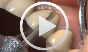
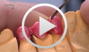
Gap Closure with a Minor Incisal Edge Restauration
Klaiber, BerndProcedure: - Incisal edge restoration at Tooth 11 - Sketch prior to widening procedure in anterior teeth region - Placement and shaping of matrix band retainer used for tooth widening - Application of composite material and spreading with small Heidemann spatula to establish stable and broad approximal contact - Application of dentin and enamel composite - Shaping and completing; positioning of the lateral edge lines; shaping the interincisal triangle. - Creation of an invisible transition, composite/enamel, with scalpel #15 - Polishing Materials Adhesive: Optibond Fl (Kerr) Composite: Enamel HFO (Micerium) Dentin UD4, UD3,5 and UD3 Enamel GE2 Opalescence OBN Flowable Composite: Tetric Flow A4 (Vivadent) Temporary Composite for shaping of matrix band retainer SystepOnlay (Vivadent) -
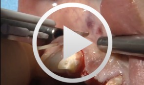
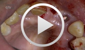
The Carnevale Technique: Hemisection/Trisection of Molars with Furcation Involvement
Hürzeler, Markus B.Procedure: - Apical Flap Repositioning - Trisection of the Upper Molar - Extraction of the Distobuccal Root - Tangential Preparation of the Abutment Teeth - Temporary Relining Contents: Molars with furcation involvement have a shorter long-term prognosis for tooth retention than single-rooted teeth. Apart from replacing these teeth with implants, they can also be treated by hemisection or trisection with the goal of eliminating furcation and creating single-root conditions. Studies on the long-term stability of teeth treated by hemisection / trisection show mixed results. Some investigators have found failure rates of up to 40 percent. In Gianfranco Carnevale's group, on the other hand, success rates of more than 90 percent have been reported for a 10-year follow-up period. Pretreatment: Initial work on the abutment teeth, which had grade II-III furcation involvement, was done six to eight weeks after conservative periodontal therapy in preparation for placement of the long-term temporary. Preparation was done tangentially, up to the bone level, to spare as much dental hard tissue as possible while eliminating all recesses and root concavities. A metal-reinforced long-term temporary was used to splint the prepared teeth. Endodontic treatment of the affected teeth was subsequently performed. Surgical procedure: Apical displacement of the gingiva around the involved teeth was done before incision. A mucosal fl ap was created on the buccal and palatal sides. Trisection of the teeth was then performed. After dissecting and excising the distobuccal root, intraoperative preparation of the abutment teeth was carried out. Temporary relining was another important step, the objective of which was to splint the root and to prevent tilting. Further treatment: Impressions for the defi nitive restoration were made six months postoperative. The restoration margins of the tangential preparation were defined using a master model. The definitive reconstruction had fine metal margins. -
Zirconia Ceramic Restorations Vennered Using the Overpress Technique, part 2
Bußmeier, UweMaterials Checklist: Zeno Tec System Wieland Software: Dental Designer by 3shape Milling unit: Wieland 4030 Zirconia: ZENO® Zr; zirconia staining: Zircolor Investment material: PressX Zr Investment Press: IMAGINE Press with disposable plungers Ingots: PressXZR SHO-3; veneering ceramic: ZIROX Glaze: PressXZR Body Stain A3 / Glaze -
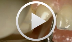
Restoration of a Class IV defect in the anterior segment
Hugo, Burkard -
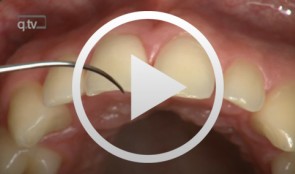
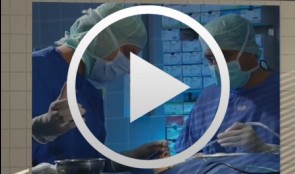
Ridge augmentation in the periodontally involved dentition
Windisch, PéterContents: - Periodontal regeneration and alveolar -ridge augmentation using a connectivetissue graft - Implant insertion and augmentation - Implant re-entry and prosthetics Materials Checklist Emdogain, Bio-Oss, BioGide, Block fixating screw for autologous bone cylinder, 4/0 and 5/0 sutures, Resolut membrane Titanium pins, Autologous bone chips, 2 Replace Groovy Tapered 4, 3x13 mm implants -
Operative Therapiekonzepte zur Entfernung retinierter unterer Weisheitszähne
Schultze-Mosgau, Stefan / Neukam, Friedrich Wilhelm / Basting, GerdGliederung: - Schematische und röntgenologische Demonstration unterschiedlicher Retentionsformen unterer, retinierter Weisheitszähne, Darstellung der Indikation zur Entfernung - Erstellung der Behandlungsunterlagen, Aufklärungsgespräche mit Darstellung der Komplikationen - Demonstration des operativen Vorgehens: Lokalanästhesie, Schnittführung, Schutz des N. lingualis, Osteotomie, Nahttechnik. Die Entfernung unterer, retinierter Weisheitszähne gehört zu den häufigsten dento-alveolär-chirurgischen Eingriffen. Durch die enge anatomische Lagebeziehung zu den benachbarten Zähnen und dem N. alveolaris inferior besteht bei der operativen Entfernung die Gefahr einer Schädigung der umgebenden Strukturen. Die Kenntnis der verschiedenen Retentionsformen und eine geeignete, atraumatische Operationstechnik ist für eine komplikationslose Entfernung von Bedeutung. Nach der Leitungsanästhesie des N. alveolaris inferior und des N. buccalis wird die Schnittführung so gewählt, dass ein vestibulär gestielter Mukoperiostlappen gehoben werden kann. Nach dem lingualen, subperiostalen Einführen eines Raspatoriums zum Schutz des N. lingualis wird durch die bukkale Osteomie mit kugelförmigen Hartmetallfräsen der Weisheitszahn bis zu seiner größten Zirkumferenz freigelegt und durch vorsichtige Luxationsbewegungen entfernt. -
Flap surgery using a modified papilla preservation technique
Salvi, Giovanni E.Outline - Introduction: Patient history, clinical findings, diagnoses, etiology, prognosis of individual teeth - Treatment planning for phases - Modified papilla preservation technique (MPPT) - Simplified papilla preservation technique (SPPT) -Clinical situation 6 months postoperatively -
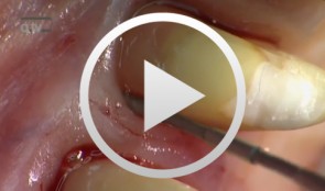
Treatment of a palatal class II furcation
Marggraf, ErwinOutline: - Reflecting a flap - Cleaning. - Fraction 3 - Fraction 2 - Fraction 1 - Wound closure List of materials All materials required for producing PRGF (BTI Germany) Bone replacement materials Geistlich Biomaterials Surgical instruments, Aesculap Suture materials, Ethicon