


-
- English
- Deutsch
- Spanish
-
- General Diagnostics
- Treatment
- Tools/Materials
- Basic/General Knowledge
- Research
- Events
- Organisations
-
-
-
-
-
-

-

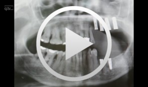
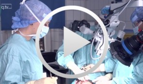
Implant placement in the anterior mandible with bone augmentation
Hürzeler, Markus B. -
Vestibuloplasty and mucosal graft in implant site re-entry following maxillary reconstruction
Schultze-Mosgau, Stefan -
-
-
-
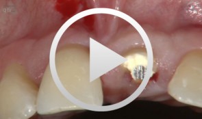
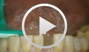
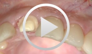
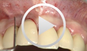
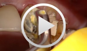
Immediate implant placement at site 21 with combination-graft closure
Iglhaut, Gerhard M. -
3D planning and template-guided implant insertion in the edentulous jaw
Kirsch, Axel / Ackermann, Karl-Ludwig / Neuendorff, GerhardOutline: - Surgical procedures for anchoring a diagnostic guide - Inserting four provisional implants - Impression and bite registration, fabrication of the master cast - Tooth-setup for the temporary restoration - Registering the setup in a silicone index - Duplicating the setup in radiopaque resin for CT imaging - Implant planning using a 3D record of the CT image - Fabricating a transfer template based on the CAD treatment plan using the CAMLOG® Guide System - Inserting the guiding sleeves into the template - Fabricating the final restoration prior to inserting the implants - Vario SR abutments with Vario SR titanium copings for a passive fit - Fabricating a cast titanium framework to reinforce the restoration - Surgical procedures demonstrating the definitive implant insertion - Insertion of six implants for immediate loading - Providing a controlled-clearance fit between the implants and the denture base -
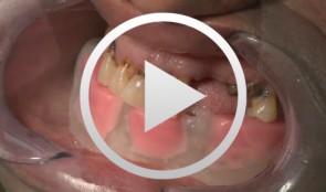
Implant-supported removable restorations in the edentulous jaw
Wolfart, Stefan / Weber, VolkerOutline: - Patient presentation, impression, comprehensive jaw relation records - Wax-up, Fabrication of the provisional restoration - Fabrication of a DVT based planning and drilling template - Surgical procedures for inserting four implants in the mandible - Suturing and relining of the existing denture as a provisional restoration - After 12 weeks: Reentry and insertion of healing abutments - Harvesting a free gingiva graft to extend the attached gingiva - Verifying implant stability using Periotest - Reworking the existing denture to fit on the healing abutments - Impressioning with custom tray (pick-up technique) - Demonstrating the line finder to transfer face axis - Fabricating the three restorations with Locator® attachments, electroplated double crown, precision-milled bar - The matrix and retention parts of the Locator® system, transferring the Locator® abutments to the implants - Fabricating the electroplated copings, intraoral adhesively connecting the electroplated copings to the cast framework (passive fit), finishing and delivery - Removable restoration on a custom-milled bar, clinical and laboratory workflow, delivery - Maintaining implant-supported restorations -
Microsurgical lateral sinus floor elevation (LSFE)
Nölken, RobertOutline: - Incision - Flap mobilization - Lateral sinus fenestration - Elevation of the Schneiderian membrane - Implant bed preparation - Bone chip harvesting at the mandibular angle - Filling of sinus lift lumen with autologous bone chips - Implant insertion - Covering the lateral sinus cavity with collagen membrane - Wound closure List of materials - Zeiss Pro Dent microscope with beam splitter and Panasonic 3 CCD camera - Scalpel holder (Ustomed) with Swann-Morton blades 15C and 12D - Narrow rasp (Hu-Friedy) - Micro-vacuum (Luer Lock Suction Tip, American Dental Systems) - Disposable vacuum tube set (Bexamed) - Disposable draping, Lindau (Aescologic) - Piezosurgery with diamond ball (Mectron) - Microforceps (Hu-Friedy) - Excavator (Martin) - Periodontometer, 1-mm gradation (Hu-Friedy) - OsseoSpeed implant set, Dentsply Implants: Marking drill; Twist drill, 2 mm; Depth gauge; Pilot drill, 2/3.2 mm; Twist drill, 3.2 mm; Tapered drill, 3.2/5 mm; OsseoSpeed TX implant, 5.0 × 11 mm; Closure screw, 4.5/5 mm - Columbia curette (Ustomed) - Micross scraper (Meta) - Needle holder (Ustomed) - Langenbeck wound retractor (Ustomed) - Kelly scissors (Ustomed) - Buchanan endodontic hand plugger (American Dental Systems) - Resorbable collagen membrane (Resodont, Resorba) - Ethilon 5-0 FS-3 (Ethicon) - Prolene 6-0 DA-2 (Ethicon) -
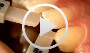
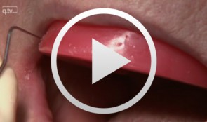
Composite Restoration in Anterior Teeth
Hugo, BurkardProcedure: - Introduction with control of the Gap Width and Colour Selection - Preparation of the Dental Surface (application of the Matrix) - Gap Closure, Final Polish Contents The present case shows the gap closure at tooth 22 with a direct adhesive technique. The gap closure is done here after the orthodontic treatment of a central diastema. A special matrix technique is used to allow a perfect design of the aproximal surfaces and the creation of the aproximal contact points. During the colour selection the dentin and enamel colours are chosen very carefully, to allow a good esthetic result and different composites are used to achieve a natural aspect of the tooth.