-
- English
- Deutsch
- Spanish
-
- General Diagnostics
- Treatment
- Tools/Materials
- Basic/General Knowledge
- Research
- Events
- Organisations
-
-
-
-
-
-

-

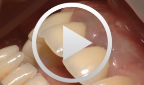
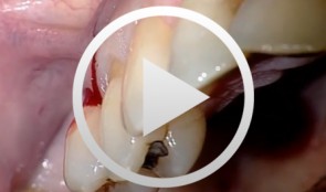
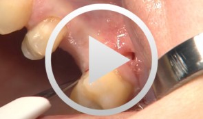
Regenerative Procedures for Optimized Esthetics at Tooth 11
Schlee, MarkusContents: - Exploration - Incision and Flap Mobilization - Palatal Flap Preservation with Interdental Tissue Preservation - Detoxification and Concrement Removal at 11 - Harvesting of Autogenous Bone Chips from the Spina Nasalis - Conditioning of the Root Surface with EDTA-Gel - Application of Emdogain and Filling of the Bone Defect - Wound Closure Synopsis After Finishing the Initial Treatment for Aggressive Periodontitis, Regenerative Treatment of a Tunnel-Shaped Pocket at Tooth 11 was attempted. Rotation and Crowding of the Buccally Inclined Tooth represented a favorable Etiological Factor. The patient did not wish to receive Orthodontic Treatment to eliminate this Causal Factor after Completion of Primary Treatment. Treatment was therefore limited to the Surgical Regeneration Attempt. The Interdental Space was larger than 3 mm and the Bone Pocket was a mostly Three-Walled Structure, so the Chances of Success were considered to be good. Exploration was first performed to identify the Course of the Defect Margins. Exact knowledge of the Bone Anatomy in all three Planes is essential to successful Incision Planning. A Tunnel-Shaped Defect delimited by Bone in the Region of Tooth 11 with good chances of Periodontal Regeneration was found. A major Challenge of this Procedure is the need to keep the Defect completely covered with Soft Tissue throughout the Healing Process. Cortellini's Papilla Preservation Technique was used for this Purpose. After Incision and Flap Mobilization, it became evident that the Defect only had two Walls in the Coronal Region and that Bone was lacking in the Buccal Region. According to the current Data on Periodontal Regeneration, the Attachment Gain achieved using an Enamel Matrix Protein (Emdogain®) alone can be just as good as that achieved using Emdogain and Bone Graft Material combined. Still, we elected to use a Combination Technique in the Present Case because it provides better Papillary Support. The Graft Material consisted of Autogenous Bone Chips from the Spina Nasalis, which can easily be harvested by Means of the Piezo Technique After gentle Detoxification, the Root Surface was treated with Emdogain. The Defect was then filled with Autogenous Bone Chips and closed by Microsurgical Suture Techniques. Six months after Surgery, Partial Regeneration of the Papilla can be seen. -
Immediate placement of a NobleActive implant in a patient with a pronounced hard-tissue and soft-tissue defect
Nölken, RobertOutline - Modified tubed flap - NobelActive implant placement - Flapless facial bone augmentation - Immediate provisionalization Materials Checklist: NobelActive™ Surgery Kit Twist drill ø 2, 7-15 mm Twist drill ø 2, 10-18 mm Twist step drill ø 2.4/2.8, 7-15 mm Twist step drill ø 2.4/2.8, 10-18 mm Twist step drill ø 3.2/3.6, 7-15 mm Twist step drill ø 3.2/3.6, 10-18 mm Surgical driver NobelActive™ Man torque wrench, surgical NobelActive™ Internal RP implant Procera® esthetic abutment, NobelActive™ Internal Implant replica, NobelActive™ Internal RP Impression coping, open tray, NobelActive™ Internal RP Protect analog, NobelActive™ Internal. -
Augmentation at site 16 using the SonicWeld Rx System
Iglhaut, Gerhard M. -
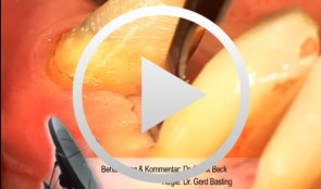
Piezo Surgery Technique for Alveolar Ridge Augmentation with Sinus Lift
Schlee, MarkusOverview: - Periodontal and implant planning - Treatment of a horizontal and vertical bone defect - discussion of the literature - Clinical implementation of bone augmentation using a bone block and treatment of a vertical pocket with Emdogain - Sinus lift in combination with alveolar ridge augmentation and horizontal expansion of the alveolar process; orthodontic straightening of a molar tooth Contents: This contribution illustrates a complex periodontal-implantological case, from treatment planning to clinical implementation. It details the transplantation of two bone blocks from the linea obliqua of the angle of the jaw to the anterior front tooth region, the treatment of a vertical bone pocket with Emdogain, the straightening of a molar tooth using orthodontic mini-implants, and a sinus lift together with alveolar ridge augmentation in the maxillary region using a piezo surgical technique. -
Minimally Invasive Implant Surgery based on Three-Dimensional CT Treatment Planning for Total Rehabilitation
Beck, FrankProcedure: - Incision Technique 36, 44 - Gentle Flap Mobilization - Pilot Hole Preparation using a CT Template - Sequential Preparation and Implantation - Bone Removal and Augmentation - Wound Closure Contents: Systematic Total Rehabilitation is very challenging, especially in patients with Periodontal Disease with Loss of Supporting Structures. Precise Treatment Planning is essential. The Treatment of Periodontal Disease begins after Conservative Pretreatment. It is not possible to predict the Soft Tissue Esthetic Outcome before the Healing Process is completed. We selected Endosteal Implants for the Augmentation of Lost Support Zones in this Atrophic Mandible. A Three-Dimensional Analysis was performed as the Basis for Navigated Implantation. After Completion of the Periodontal Treatment and Implantation, the patient must wait for approximately 6 months for the Completion of the Healing Process before Prosthetic Reconstruction Procedures can be initiated. -
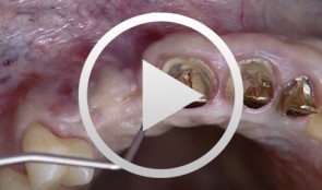

Implantation with Simultaneous Augmentation
Grunder, UeliProcedure: - Case evaluation - Incision technique - Implant placement - Membrane adjustment and fixation - Introduction of replacement material - Flap mobilization - Suture technique Contents: Implantation was desired for replacement of a missing upper canine tooth and the adjacent lateral incisor tooth. The initial case evaluation revealed a relatively narrow gap between these two teeth in addition to extensive hard and soft-tissue defects. We selected an incision technique that made it possible to do the augmentation work yet subsequently achieve a tension-free flap closure. Since the bony defect was large while the available space was limited, we had to go for the best possible compromise in regard to implant insertion. After the implants had been inserted, augmentation was carried out using a non-absorbable, titanium-reinforced membrane, bone replacement material, and an absorbable membrane. Extreme flap mobilization was needed to achieve flap closure. An optimal suture technique was used to complete the surgery. -
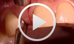
Single Tooth Replacementwith Implants in the Esthetic Region
Yüksel, OrcanProcedure: Delayed loading of dental implants in the esthetic zone of the maxilla. Guided bone regeneration (GBR) for compensatory augmentation with subsequent exposure after healing. Contents: Soft and hard tissue loss leads to esthetic problems, even in patients with successful implant osseointegration. Delayed loading of dental implants in the esthetic zone is frequently indicated in dental practice, for example, in patients with congenital absence of the lateral incisors. Dental implants can solve this problem. Depending on the extent of hard tissue loss, it may be necessary to perform guided bone regeneration (GBR) in order to achieve better esthetic results. Depending on the degree of atrophy, GBR may be performed before or simultaneous with implantation. Correct implant placement is essential to achieving the end goal: an esthetically pleasing and natural result. In this film and in the presentation, we will demonstrate in detail the procedure for placing implants in this esthetically sensitive region. In addition, we will demonstrate a method of impression-taking that provides dental laboratories a better foundation for achieving excellent esthetic results. -

Ballon Lift Control - A life report
Heuckmann, Karl-Heinz -
Internal Sinus Lift with the Lift Control System
Streckbein, Roland -