


-
- English
- Deutsch
- Spanish
-
- General Diagnostics
- Treatment
- Tools/Materials
- Basic/General Knowledge
- Research
- Events
- Organisations
-
-
-
-
-
-

-

Procedings of the 10th European Workshop in Periodontology of the EFP and Osteology Foundation - original publication open access
EFP and Osteology FoundationSharing the same goals made teaming up an easy task for the EFP and Osteology Foundation when they jointly steered the 10th European Workshop in Periodontology to success in November 2013 in La Granja de San Ildefonso, Spain. The proceedings of this workshop are available with free access in a special supplement of the Journal of Clinical Periodontology (JCP). -
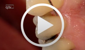
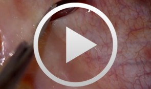
Veneer preparation in cases with wedge-shaped defects
Kopsahilis, NikolaosContents: - Medical history and diagnosis - Dental status, periodontal status, functional status - Periodontal therapy, functional therapy - Conservative treatment - Restoration (a: posterior; b: anterior) Materials Checklist Sutures: - Gingi-Plain - Z-Twist - Non-impregnated (Gingi-Pak) Impression materials: - Impregum Penta Soft (3M ESPE) - Permadyne Garant 2:1 (3M ESPE) -
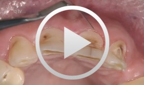
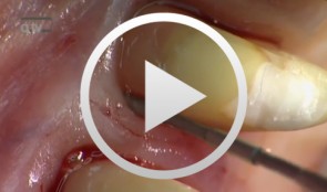
Periodontal regeneration at teeth 21 and 23 using EMD and cortical bone chips
Topoll, Heinz H.Contents: - Incisions using a microsurgical scalpel - Reflecting a buccal flap - Preparing papillary flaps using a microsurgical scalpel - Lifting off of the papillary flaps using a papillary elevator - Removing the granulation tissue using an ultrasound scaler - Cleaning the dental roots using manual instruments - Trying to dental root - Applying Emdogain - Mixing Bio-Oss and Emdogain - Introducing the Bio-Oss into both bone defects - Microsurgical suturing Materials Checklist: Cheek retractor Microsurgical scalpel blade holder Microsurgical scalpel blade Soniflex tips Bone rest Castroviejo microsurgical needle holder Suturing scissors Dental tweezers Microsurgical tweezers Monofilament suturing material 6/0 Seralene Pref gel Emdogain Bio-Oss -
Ridge augmentation in the periodontally involved dentition
Windisch, PéterContents: - Periodontal regeneration and alveolar -ridge augmentation using a connectivetissue graft - Implant insertion and augmentation - Implant re-entry and prosthetics Materials Checklist Emdogain, Bio-Oss, BioGide, Block fixating screw for autologous bone cylinder, 4/0 and 5/0 sutures, Resolut membrane Titanium pins, Autologous bone chips, 2 Replace Groovy Tapered 4, 3x13 mm implants -
Regenerative Treatment on Tooth 14 und 24
Eickholz, PeterProcedure: - Incision - Flap Design - Removal of the granulation tissue - Application of the PrefGel on the root surface - Application of the Enamel -Matrix -Protein (Emdogain) - Suture (Offset-Suture) - Identical procedure on the opposite side (1st quadrant) Materials: Retractor Micro Surgical Scalpel Handle Mini Scalpel Blades 4 x Gracey Curettes Periosteal Trombelli Periosteal Prichard Microsurgical Needle Holder Castroviejo Scissors Tweezers Microsurgical Tweezers Gore Tex CV-5 Sutures Gore Tex CV-6 Sutures Emdogain 0,7 ml PrefGel -
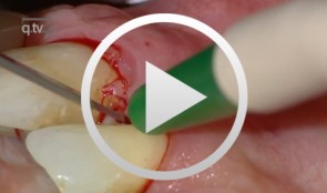
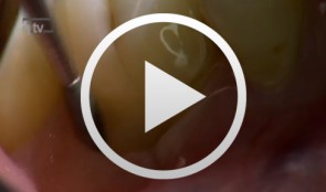
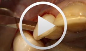
The Carnevale Technique: Hemisection/Trisection of Molars with Furcation Involvement
Hürzeler, Markus B.Procedure: - Apical Flap Repositioning - Trisection of the Upper Molar - Extraction of the Distobuccal Root - Tangential Preparation of the Abutment Teeth - Temporary Relining Contents: Molars with furcation involvement have a shorter long-term prognosis for tooth retention than single-rooted teeth. Apart from replacing these teeth with implants, they can also be treated by hemisection or trisection with the goal of eliminating furcation and creating single-root conditions. Studies on the long-term stability of teeth treated by hemisection / trisection show mixed results. Some investigators have found failure rates of up to 40 percent. In Gianfranco Carnevale's group, on the other hand, success rates of more than 90 percent have been reported for a 10-year follow-up period. Pretreatment: Initial work on the abutment teeth, which had grade II-III furcation involvement, was done six to eight weeks after conservative periodontal therapy in preparation for placement of the long-term temporary. Preparation was done tangentially, up to the bone level, to spare as much dental hard tissue as possible while eliminating all recesses and root concavities. A metal-reinforced long-term temporary was used to splint the prepared teeth. Endodontic treatment of the affected teeth was subsequently performed. Surgical procedure: Apical displacement of the gingiva around the involved teeth was done before incision. A mucosal fl ap was created on the buccal and palatal sides. Trisection of the teeth was then performed. After dissecting and excising the distobuccal root, intraoperative preparation of the abutment teeth was carried out. Temporary relining was another important step, the objective of which was to splint the root and to prevent tilting. Further treatment: Impressions for the defi nitive restoration were made six months postoperative. The restoration margins of the tangential preparation were defined using a master model. The definitive reconstruction had fine metal margins. -
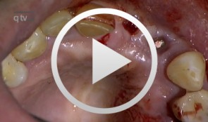
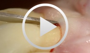
Regenerative Treatment of Class II Mandibular Furcation Defects
Heinz, BerndProcedure Case description: -Class II furcation defect at teeth 46 and 47 and gingival recessions at teeth 43 and 44 - Root planing using PerioSet - Incision technique - Cleaning furcation defect at tooth 46 - Pref Gel application, rinsing and Emdogain application - Insertion of Bio-Oss into the furcation space with an amalgam plugger after hydration - Condensation of the bone replacement material and application of an absorbable membrane (Bio-Gide) - Atraumatic suture closure using 6/0 Seralene Contents: This video demonstration shows the simultaneous treatment of recessions at teeth 43 and 44 and of class II furcation defects at teeth 46 and 47. After a brief case description, root planning is done using PerioSet. Next, an incision is made and the furcation defects are very carefully cleaned using hand instruments and ultrasonic scalers (Soniflex). The cleaned root surfaces and furcation defects are conditioned with Pref Gel (Straumann) for two minutes. The objective of conditioning is to remove the smear layer, to open the dentine tubules, and to enable surface demineralization. Moreover, this measure serves to optimize the contact between Emdogain and the root surface. After two minutes, the EDTA suspension is removed using physiological saline solution or water spray. Immediately afterwards, Emdogain is applied to the blood and saliva-free root surface. This procedure was also used to treat the furcation defect at tooth 47. Regenerative treatment of tooth 46 was performed since that tooth had a very extensive furcation defect. The defect was filled with Bio-Oss, which was applied using an amalgam plugger. Absorbable Bio-Gide was used for coverage of the furcation entrance. Finally, the wound was closed using loop sutures and single interrupted sutures. -
Flap designs for Interdental Tissue Preservation in Periodontal Therapy
Salvi, Giovanni E.Procedure: - Introduction: History -Taking, Examination, Diagnosis, Etiology, Prognosis for Individual Teeth - Four - Phase Treatment Sequence - Modified Papilla Preservation Technique(MPPT) - Simplified Papilla Preservation Technique (SPPT) - Findings 6 months after Surgery Synopsis The Modified and Simplified Papilla Preservation Techniques for conservation of interproximal papillary tissue were designed to provide access to deep and narrow bony defects to enable regenerative periodontal treatment. The Modified Papilla Preservation Technique (MPPT) was designed to ensure tension-free primary closure via barrier membranes in patients with small interdental spaces. The Simplified Papilla Preservation Technique(SPPT) is used to gain access to narrow interdental spaces ( < 2mm) and to deep defects in the lateral tooth region. Apart from preserving primary wound closure in the interdental space, the two techniques also serve to keep the membrane from collapsing into the bony defect. Both MPPT and SPPT employ special suture techniques to ensure tension-free primary closure of the interdental space. This video clip also serves to demonstrate that the two techniques of interproximal tissue preservation can also be used for periodontal interventions without regenerative measures. -
Regenerative Procedures for Optimized Esthetics at Tooth 11
Schlee, MarkusContents: - Exploration - Incision and Flap Mobilization - Palatal Flap Preservation with Interdental Tissue Preservation - Detoxification and Concrement Removal at 11 - Harvesting of Autogenous Bone Chips from the Spina Nasalis - Conditioning of the Root Surface with EDTA-Gel - Application of Emdogain and Filling of the Bone Defect - Wound Closure Synopsis After Finishing the Initial Treatment for Aggressive Periodontitis, Regenerative Treatment of a Tunnel-Shaped Pocket at Tooth 11 was attempted. Rotation and Crowding of the Buccally Inclined Tooth represented a favorable Etiological Factor. The patient did not wish to receive Orthodontic Treatment to eliminate this Causal Factor after Completion of Primary Treatment. Treatment was therefore limited to the Surgical Regeneration Attempt. The Interdental Space was larger than 3 mm and the Bone Pocket was a mostly Three-Walled Structure, so the Chances of Success were considered to be good. Exploration was first performed to identify the Course of the Defect Margins. Exact knowledge of the Bone Anatomy in all three Planes is essential to successful Incision Planning. A Tunnel-Shaped Defect delimited by Bone in the Region of Tooth 11 with good chances of Periodontal Regeneration was found. A major Challenge of this Procedure is the need to keep the Defect completely covered with Soft Tissue throughout the Healing Process. Cortellini's Papilla Preservation Technique was used for this Purpose. After Incision and Flap Mobilization, it became evident that the Defect only had two Walls in the Coronal Region and that Bone was lacking in the Buccal Region. According to the current Data on Periodontal Regeneration, the Attachment Gain achieved using an Enamel Matrix Protein (Emdogain®) alone can be just as good as that achieved using Emdogain and Bone Graft Material combined. Still, we elected to use a Combination Technique in the Present Case because it provides better Papillary Support. The Graft Material consisted of Autogenous Bone Chips from the Spina Nasalis, which can easily be harvested by Means of the Piezo Technique After gentle Detoxification, the Root Surface was treated with Emdogain. The Defect was then filled with Autogenous Bone Chips and closed by Microsurgical Suture Techniques. Six months after Surgery, Partial Regeneration of the Papilla can be seen. -
Regenerative Measures for Osseous Defect Repair and Optimal Esthetics
Sculean, AntonProcedure: Theoretical Part: - Adult male with a deep and broad intraosseous bone defect located on tooth #13 - The indication for modified papilla preservation in the scope of regenerative therapy was established based on the width of the diastema - Regenerative periodontal therapy with Emdogain and a Bio-Oss® cancellous bone graft - Emdogain is applied to the root surface to stimulate regeneration of periodontal structures - To prevent graft collapse and to minimize the risk of development of too large a recession in this esthetically important region, the defect was filled with Bio-Oss® cancellous bone material Practical Part: - The papilla preservation technique was performed using microsurgical instruments - The root surface area was conditioned with 24% EDTA for ca. 2 minutes - Emdogain was applied to the root surface - The defect was filled with the Emdogain/Bio-Oss® mixture - The wound was closed with two mattress sutures one horizontal mattress suture to secure the graft in place, and a second modified vertical mattress suture to tightly close the papilla - A 5-0 suture was used for the horizontal mattress suture, and a 6-0 monofilament was used for the vertical mattress suture - Postoperative care entailed rinsing the wound twice daily for 4 weeks with 0.2% chlorhexidine and ibuprofen analgesia on the first few days after surgery Contents: The patient's jaw displayed a generalized loss of clinical attachment and alveolar bone. His general history was unremarkable; the patient was a non-smoker. Microbiological tests showed large numbers of Actinobacillus actinomycetemcomitans and Porphyromonas gingivalis. The diagnosis was "generalized aggressive periodontitis". After four months of initial therapy consisting of antibiotic combination therapy (amoxicillin + metronidazole), intraoral radiographs showed a deep and wide intraosseous bone defect located mesial and palatal to tooth #13. To preserve this strategically important tooth we opted to perform regenerative therapy with Emdogain and Bio-Oss cancellous bone material. Ten months after regenerative periodontal therapy, the probing depth had decreased by 7 mm, and 5-6 mm of clinical attachment had been gained. At this time, the probing depth was 2-3 mm and intraoral radiographs showed near-complete filling of the osseous defect.