


-
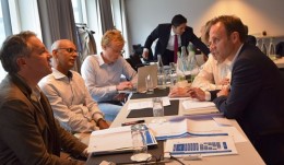

Osteology Expert Meeting
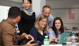
Internationally renowned experts with great experience and interdisciplinary expertise in simulation gathered in Zurich for the 1-day Expert Meeting to discuss the topic and share their expertise in this field with theFoundation Boardand representatives of theExpert Councilof the Osteology Foundation attending the meeting. The Osteology Foundation organizes an Expert Meeting every year to discuss a topic either of high relevance for science and practice in oral regeneration or of importance for the future development of the Foundation and its activities or the dental field in general, as was the case at the 2015 meeting. The aim of the Expert Meeting on simulation in training and education was to evaluate the state of the art in other disciplines, to find out which new possibilities current developments and technologies allow, and the consequences for the Osteology Foundation on the one hand, but also for the whole field of professional training and education in dentistry, on the other. External speakers and experts invited to the 2015 meeting wereProfessor Amitai Ziv, Israel,Professor Margaret Cox, UK,Professor Gabor Szekely, Switzerland,Dr David Joseph, France, andArnaud Cosson, France. In addition,Dr Stefan Tuchschmid, Switzerland, joined for the discussions.Dr Franck Renouard, France, Board Member of the Osteology Foundation, gave an introduction to the subject area, emphasising the overall importance of training. To err is human Professor Amitai Ziv, Founder and Director of theIsrael Centre of Medical Simulation(MSR), and also a veteran combat pilot and trainer from the Israeli Air Force gave a truly eye-opening talk, demonstrating different applications of simulation in training and the impact they can have in the practice. As a medical professional, he demonstrated the crucial importance of medical simulation, but also being a pilot, he placed a special focus on lessons learned from the aviation field. In late 1999, the important report on patient safety To Err is Human was issued by the US National Institute of Medicine, Ziv told the audience. It indicated that health care is far less safe than it should be, and that deaths due to medical errors in the US number almost 100,000 annually. The report triggered a massive response, and simulation in health care has grown rapidly ever since. Today simulation is used to reproduce real patient experiences and encounters using guided and controlled simulation-based scenarios, Ziv continued. It offers a safe and mistake-forgiving environment where trainees can learn from their errors without risking harm to real patients. Simulation provides a hands-on empirical educational modality, enabling controlled proactive exposure of trainees to both regular and complex, uncommon clinical scenarios. It also provides an opportunity for team training, which is important for patient safety, but is seldom addressed in traditional medical education. Another important benefit is the reproducible, standardized, objective setting it provides for assessment purposes, Ziv added. He demonstrated the impact of the training by showing examples from his institute, the MSR, which operates as a national simulation centre conducting mandatory training and assessment programs in multiple clinical fields. At MSR they use actors, models and virtual techniques to simulate a huge variety of training situations. Rapid increase in computational power Professor Gabor Szekelyin his presentation provided an introduction into the technical aspects of simulation for medical training, what is possible now, and what may be possible in the future. Szekely is head of theMedical Image Analysis and Visualization Groupat the Swiss Federal Institute (ETH) in Zurich and is also Chairman of the Swiss Institute of Computer Aided Surgery. The rapid increase in computational power available, as well as current capabilities in computer graphics and virtual reality, enable the use of advanced, complex simulation systems, Szekely explained. As a result, simulation is set to become a useful tool in everyday clinical practice, efficiently assisting surgeons in dealing with difficulties in the operating room. Szekely presented examples for the development of surgical simulators, and the major of technological components necessary for their development. Medical simulation mainly concentrates on minimally invasive procedures, he explained, because there are still many obstacles towards creating realistic haptic training simulators for open surgery. Implementation in the dental field Professor Margaret Cox, Professor of Information Technology in Education at Kings College London, who has developed educational software for more than 40 years, presented a haptic device that she and her working group have developed for dental education. The aim of thehapTEL projectis to develop and evaluate a haptic environment for preparing cavities in a virtual tooth. The outcome of the project included setting up 14 fully functional hapTel curriculum workstations, which have been integrated into the dental undergraduate curriculum for six years, explained Cox. Educational evaluation results showed that the students who learn on the hapTEL workstations performed just as well as those learning in the traditional Phantom Head laboratory, with the added advantage of being able to review their progress at any time and seeing exactly how well they are doing. Cox also reported that since July 2011 the work of the project has been extended and modified hapTEL work-stations now enable students to learn how to give injections. Simulation of implant placement Another example where simulation is used, was presented byDr David Josephfrom the University of Nancy. He presented initial results obtained with a haptic simulator (VirTeaSy) that has been used to train and evaluate students in implantology. They observed improved performance in students who trained on the simulator with results close to those of experienced operators (Joseph et al. Biomed Res Int 2014). Implant placement simulators are now commercially available, said Arnaud Cosson, CEO ofHRV, the French company producing and marketing the simulators in combination with a 3D realistic immersive environment and the corresponding software that offers about 70 exercises in which realistic clinical situations are simulated. Experience with surgical simulators The participants of the meeting had the opportunity to try out an arthroscopy simulator used in orthopaedics. The highly realistic haptic simulator presented by the Zurich-based companyVirtaMed demonstrated the technical possibilities in the field of simulation. Drawbacks in current education During workshops in the afternoon, the Osteology Foundation Board and members of the Osteology Expert Council took the chance to discuss the topic in small groups with the invited experts. The discussions revealed drawbacks and deficiencies in current training at universities, but also in continuing education for practitioners in the dental field. Both recognition of the importance of simulation in dentistry, as well as the current technologies is still not common, the experts agreed. In discussions, the participants of the Osteology Expert Meeting came to the conclusion that various types of mistakes can be avoided through more extensive or better training. Three type of mistakes were identified: mistakes caused by lack of knowledge, mistakes arising from lack of experience, and mistakes relating to attitude. As general problems that exist in training students, the following points were listed: the availability and heterogeneity of cases, limited personal resources, and the impossibility of re-doing procedures from scratch in case of mistakes. In addition, the training of soft skills and reflection on mistakes is lacking in student education, and interdisciplinary education is often completely neglected. The participants of the Osteology Expert Meeting in Zurich agreed that there is a great need for simulation in dental education in two different areas of expertise: Firstly, for the training of processes, simulating stress, and using soft skills in different clinical situations. And secondly, realistic haptic training of different procedures and practical skills, where simulation is to be used in combination with existing and well-established techniques. Consequences for training in dentistry? After the presentations and discussions with the experts, the Osteology Board and Expert Council arrived at the conclusion that simulation in training is certainly something that should not be neglected. Deficiencies in current training and education in dentistry exist and current methodology can be improved. How these deficiencies can be eliminated and the role the Osteology Foundation could play, remains open and has to be discussed further. The Osteology Foundation thanks all experts, speakers and contributors to the Expert Meeting for sharing their knowledge and opening up new perspectives for training and education in dentistry. -

Osteology Education Grants 2016 – apply now!
Apply now! Application deadline for 2016 Education Grants is 1 December 2015. Applicants will be notified whether their application was accepted by 15 February 2016. Further information about the Osteology Research Academy Courses The Osteology Foundation calls for applications for Education Grants for 2016. The Osteology Education Grants are offered to support postgraduate education in the field of oral tissue regeneration. All young clinicians and basic scientists with an academic affiliation are invited to apply. The amount of funding is 2500 Swiss francs per person for participation in theOsteology Research Academycourse in Lucerne, and 2000 Swiss francs per person for participation in the courses in Boston and Berne or Vienna (final location will be announced in the course of this month). The grants are spoken based on the review of a curriculum vitae, a publication list, motivation letter, a copy of graduation certificate and a recommendation letter. Financial needs are also taken into account. For detailed information about how to apply visit theOsteology Education Grant websiteand check out theApplication Guidelines. -
Register now for Osteology Monaco 2016
Watch the official Osteology Monaco 2016 trailer Dont miss the opportunity to attend the International Osteology Symposium from 21-23 April 2016 in Monaco. Register online and book your accommodation now on our symposium website. Over the course of the three-day symposium on the topic Learning the WHY and the HOW in regenerative therapy, top-class experts will discuss the latest clinical innovations, research results and current treatment concepts in the field of regenerative dentistry. If you are interested in specific courses and workshops, you will also be able to choose from an attractive programme with 3 Master Clinician Courses (at no additional charge) and 20 practical and theoretical workshops on the first day of the congress. -
Answers to open questions - for better treatment of patients
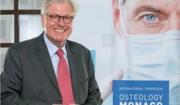
Are you interested to attend Osteology Monaco 2016? Check out the symposium website atwww.osteology-monaco.organd register now! Professor Myron Nevins, USA, is one of the Chairmen of Osteology Monaco 2016. In an interview he explained what he considers the most important topics to be addressed in the scientific programme, and why dentists should visit the international Symposium in April 2016. Professor Nevins, the Scientific Program of the 2016 Osteology Symposium with its motto Learning the WHY and the HOW in regenerative therapy addresses the many open questions that dentists still face in everyday work in the practice. Which are the most important questions in oral regeneration to be discussed in Monaco? Myron Nevins:All regenerative procedures require surgical skills, knowledge of the appropriate steps and the use of biomaterials that have proven to be successful. It is important that clinicians and practitioners not only understand the basics, but also hear about the latest developments and techniques in oral regeneration. With the International Osteology Symposium, we aim to provide this information, as well as answering as many open questions as possible. The programme covers a broad range of topics in oral regeneration, which are especially important to you? Myron Nevins:I would like to point out the two interactive sessions we have organized for Osteology Monaco. On Friday the focus will be on decision-making after tooth extraction, the second session on Saturday is about treatment of demanding gingival recessions two very important topics for practitioners. We have invited renowned experts to discuss these topics and provide answers to questions regarding indications, surgical techniques, tips and tricks. Questions can be submitted in advance, but the audience is also welcome to ask during the session. One topic of the scientific programme is teeth for a lifetime. What does this involve? Myron Nevins:Keeping ones teeth a life long means never becoming edentulous. This can be accomplished predictably with the patients natural dentition or by supplementing with implants. But in this session we will focus on the preservation of the patients own teeth, i.e. how we can preserve periodontally compromised teeth, how regenerative therapies improve tooth prognosis, as well as the advances and limitations in treating furcation defects. An extensive poster exhibition and a research forum will present the latest results in basic and clinical research. How important are these results from research for the practitioner? Myron Nevins:It is necessary to first perform pre-clinical research that can then be translated into clinical research to then be applied in practice for the patients benefit. This process takes a significant period of time and has to clear regulatory hurdles. At the Osteology Symposium in Monaco a platform is provided for researchers to present and discuss their results with other researchers on the one hand, but also to encourage the exchange between scientists and practitioners, on the other, which is important for both groups working hand-in-hand towards the same goal: to provide optimal treatment for patients. If your colleagues ask you why they should attend Osteology Monaco, what do you tell them? Myron Nevins: Dentists should attend Osteology Monaco to help provide better treatment for their patients by learning about the latest developments in oral regeneration, through discussions and interaction with their colleagues. Thank you for the interview, Professor Nevins. -
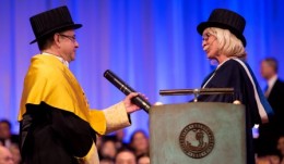
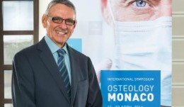
Honorary doctorate awarded to Mariano Sanz
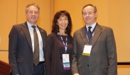
Photos: Johan Wingborg, University Gothenburg, Sahlgrenska Academy The Osteology Foundation congratulates its President, Professor Mariano Sanz, on a honorary doctorate from the University of Gothenburg, which he received for his longstanding collaboration with researchers at Sahlgrenska Academy. In May 2015 the Sahlgrenska Academy at the University of Gothenburg announced that they awarded ahonorary doctorate to Mariano Sanz,Professor of Periodontology and Dean of the Faculty of Odontology at University Complutense of Madrid, Spain, and president of the Osteology Foundation.The honorary doctorate was now conferred in a ceremony on 16 October 2015. He has published more than 200scientific articles and book chapters and is one of the most popular international lecturers on odontology, Sahlgrenska Academy said in their official announcement. Hislongstanding collaboration with researchers at Sahlgrenska Academy had a major impact on advances in knowledge about healing processes after tooth extraction. Honorary doctorates fromSahlgrenska Academyare awarded to those who have played a key role in research or education at the academy or who have otherwise promoted progress in the fields that it addresses. Together with Mariano Sanz, two other researchers were awarded:Professor Sharon Fonn from theUniversity of Witwatersrand in Johannesburg, South Africa, fora project that supports postgraduate studies in southern Africa and affords faculty members from Sahlgrenska Academy a prominent role, andProfessor Salim Yusuf,from McMaster University, Ontario, Canada, for hiscontributions to Swedish research, particularly in connection with a number of large studies concerning cardiovascular disease in low and middle income countries. More information on the website of the Sahlgrenska Academy -
Research Academy - Registration for 2016 is open now
The Osteology Research Academy goes into its sixth year. In the past five years 184 people from 39 different countries have already been educated in the Foundations intensive courses on good scientific practice and research methodology. In 2016 three courses will take place. One Core Module Course is organised in Lucerne and one in Boston. The Expert Module on Hard Tissue Research will take place in Vienna.Registration is open now. Core Module Good Research Practiceand Essentialsof Research Methodology 12-16 September 2016 Lucerne, Switzerland 24-27 October 2016 Boston, USA Expert Module Hard Tissue Research 7-9 November 2016 Vienna, Austria Financial Support Education Grants are available for participation in the Research Academy. Grants are spoken based on the review of a curriculum vitae, a publication list, motivation letter, a copy of graduation certificate and a recommendation letter. Financial needs will be taken into account. The application deadline has been extended until 15 December 2015. Apply now! -
Professor Neukam's programme highlights for Monaco 2016
The preservation of teeth is one of the most important topics for Professor Friedrich W. Neukam at the International Osteology Symposium 2016. We spoke to him in his role as the symposiums chairman on what this implies exactly and what the other important questions are which will be dealt with in Monaco in April 2016. Professor Neukam, the scientific programme of the International Osteology Symposium 2016 in Monaco will be held under the motto Learning the WHY and HOW in regenerative therapy. What are the contents that bring this motto to life? Friedrich Neukam:in our daily practice routines we need to examine all our procedures which we have applied for years - and quite successfully as we believe. The reason being, that our knowledge has changed rapidly within the space of only a few years. That is also one of the objectives of the Osteology Symposium in Monaco 2016: we want to question things and want to give dental practitioners a bench mark by reviewing known techniques in regenerative therapy, while at the same time posing the question of what is the protocol most suited to achieve a predictable treatment objective. Of course, a further objective is to present the current state of knowledge and the latest research results in the regeneration of oral tissue, to discuss these and to demonstrate their clinical implementation for dental practitioners. The programme covers a broad spectrum of topics in oral regeneration. Which are the most important for you? Friedrich Neukam:the long-term success of regenerative measures, be it to preserve teeth or for implants in healthy oral hard and soft tissue conditions. Teeth for a lifetime is a very important subject for me, to achieve the dream of preserving teeth for a lifetime if at all possible, and, of course, this also applies to implants. What are the options that techniques for bone regeneration and soft tissue regeneration offer us here in the various indications, be it in the posterior or the anterior regions - while ensuring a long-term aesthetic outcome. Of particular importance to me, is the question of which decision and treatment avenues we follow if extraction of a tooth proves necessary. Because then there are a number of options available. We need to consider the various treatment options, which is why I am looking forward to the interactive discussion on Decision making after tooth extraction. The discussion round includes experts with many years of clinical experience who will be presenting the use of regenerative techniques for preserving hard and soft tissues. You have just mentioned the topic Teeth for a lifetime. What exactly are the questions relating to this part of the programme? Friedrich Neukam:Well, as already mentioned briefly, I believe it is the wish of everyone to keep their teeth for a lifetime if possible. For me, the questions of periodontal treatment and regenerative therapy options are at the forefront here. However, what I truly look forward to, is how the HOW will be presented. For me this also means that one must receive clear information on when these measures are not effective, and I think that a crucial question will be answered: which is, when do we need to extract teeth in order to enable other treatment, such as regenerative techniques but also implant restorations in the local tissues. A major poster exhibition will also be held as part of the Osteology Monaco event, together with the Research Forum where current research results will be presented. What, in your opinion, are the most important topics which have so far been covered in research? Friedrich Neukam:if we look at the regenerative options available today, then pre-clinical research results, and of course, also clinical studies, relating to bone preservation after tooth extraction as well as the preservation and reconstruction of the soft tissue are of prime importance to me as they have a major influence on our clinical treatment modalities. If your colleagues ask you why they should participate in next years Osteology Monaco, what would you tell them? Friedrich Neukam:first of all I would not focus on the casino in Monaco, but rather I believe, that the Osteology Foundation and the international Osteology Symposium in Monaco have become the standard - a standard worldwide for regenerative measures in the oral cavity. This is where experts from across the globe meet to take positions on current topics, research results as well as clinical questions. In other words, this is a highly attractive forum with a programme which is also devoted to entirely practical aspects, namely, the WHY and the HOW in regenerative therapy. Thank you for the interview Professor Neukam. More information and registration on www.osteology-monaco.org #OsteologyMonaco -
Joint Symposium with AAP/AAP Foundation - great partnership for success
Nothing but positive feedback could be heard after the very successful symposium jointly organized by the Osteology Foundation, the AAP, and the AAP Foundation. It took place as part of the pre-programme of the AAP Annual Meeting, which was held in Orlando, Florida in November this year. With an audience of more than 500, the Symposium exceed all expectations. All seats were taken, and some late-comers even had to stand. Joan Otomo-Corgell, President of the AAP, opened the symposium, and particular welcomed over 130 third-year periodontics residents who had received scholarships from the AAP Foundation to attend the symposium. Theprogrammecovered a broad range of topics in periodontal treatment and research, exploring the current concepts and alternatives in bone regeneration, outlining the use of scaffolds and biological mediators to improve the outcomes of bone regenerative therapies, and explaining the clinical guidelines associated with current concepts and alternatives in soft tissue augmentation and periodontal regeneration. The symposium with national and international speakers also included discussion of using soft tissue substitutes to improve the outlines of soft tissue regenerative therapies. Mariano Sanzfrom Spain, current President of the Osteology Foundation (OF), andWilliam V. Giannobile(USA), Board Member of the OF, were among the presenters, as well asPamela K. McClain(USA), also Board Member of the OF and past-president of the AAP.David M. Kim(USA) andRodrigo Neiva(USA), both members of theOF Expert Council, gave presentations as well. A great addition to the programme was the presentation byMartha Somerman, Director of the National Institute of Dental and Craniofacial Research (NIDCR), who spoke about research opportunities in regenerative medicine. The organizers and everybody involved were extremely excited about the success of the symposium, and representatives of the two Foundations as well as the AAP announced to continue the collaboration, since it is a great partnership for success. More than 500 participants attended the joint symposium by the Osteology Foundation, the AAP and the AAP Foundation in Orlando, Florida, on 14 November 2015. The outstanding programme with national and international speakers focused on current technologies for hard and soft tissue oral regeneration. -
OSTEOLOGY LAUNCHES THE BOX
At the International Osteology Symposium in Monaco on 21 - 23 April 2016 the Osteology Foundation will launch its new Global Osteology Community Platform, THE BOX, which aims to provide not only tools and information, but also to link everybody in oral regeneration. An important goal for the future strategic development of the Osteology Foundation is to create a worldwide network in oral regeneration. However, it is not easy and a big challenge to link scientists and practitioners in a continuously growing global context. With this in mind, the Osteology Foundation has set up the novel Global Osteology Community Platform,THE BOX, which on the one hand provides information and tools, and on the other also allows scientists and practitioners to link up and interact worldwide, as well as also supporting all existing activities of the Osteology Foundation online. THE BOXwill be launched at Osteology Monaco 2016, and will play an important role during the symposium itself, making it also an online event by offering additional online content and benefits for participants as well as everybody else who had signed up for the online platform. THE BOX WHAT IS INSIDE? In the run-up to the congress,Hector Rios, USA, Project Leader of THE BOX, said that it was the goal of the project team to create the one place to go in oral regeneration, connecting everybody in this field, and providing new and unique benefits for scientists and practitioners. Practitioners can use a tool called the CASE BOX to document and evaluate clinical cases in oral regeneration, explained Rios. The user decides whether he wants to share his cases with his network, discuss it with a colleague or keep it private. Users can also browse the public case collection. Furthermore, THE BOX offers a novel tool for young and unexperienced scientists, the Research Wizard. It guides the user step by step through the process of setting up a research project and redirects him to relevant resources, explained Rios. Further information about protocols and scientific background is available in the online versions of the Osteology Research Guidelines. THE BOX will offer real additional benefits to the Foundations live symposia, not only during, but also after the event, said Rios. At Osteology Monaco, users will be able to access the programme online, browse all abstracts and they can also find all posters here in an online poster exhibition. The poster authors can be contacted directly, and questions to the speakers and experts will be submitted online via THE BOX during the congress. A true novel development directly linked to THE BOX is the Audience Poster Award, which will be awarded to the poster that receives the most votes online during the symposium. Rios explained further that THE BOX also provides great networking possibilities, making it possible to interact with researchers and practitioners worldwide, to extend the personal network, to find new contacts, and join discussions with experts. Furthermore, in the personally customisable part of My BOX the users find the latest news and updates from their network and everything they have saved, contributed, or where they were involved with the Osteology Foundation. Rios announced that continuous development of the platform is planned. The Foundation will create more tools and content, and also work on educational material. Rios invited every scientist and practitioner working in oral regeneration to sign up, to become part of the global Osteology community - not only for Osteology Monaco, but to help it grow and to turn it into the one place to go in oral regeneration. And the best thing, concluded Rios, is that the use of THE BOX is free. -
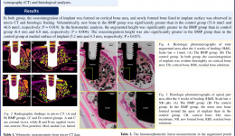
SINUS AUGMENTATION USING RECOMBINANT HUMAN BONE MORPHOGENETIC PROTEIN-2 (rhBMP-2)-LOADED SYNTHETIC BONE SUBSTITUTE WITH SIMULTANEOUS IMPLANT PLACEMENT IN RABBITS
Myung-Jae Joo (South Korea), Jae-Kook Cha, Hyun-Chang Lim, Jung-Seok Lee, Seong-Ho Choi, Ui-Won Jung Objectives: This study aimed to evaluate the effect of rhBMP-2-loaded synthetic bone substitute on implant simultaneously placed with sinus augmentation in rabbits. Methods: Following preparation of circular access windows on the maxillary sinuses, simultaneous implant (ø3x 6mm) placement with bone grafting was performed in rabbits (n=5). For the experimental group, rhBMP-2-loaded synthetic bone substitute was placed at one sinus, and saline-soaked synthetic bone substitute was placed at the other as a control. After 4 weeks of healing, block sections containing the augmented sinus and implants were obtained for micro-CT and histological analyses. Results: The volume of new bone was significantly greater in the experimental group than in the control group (p Conclusions: Within the limitations of the study, placing the implant simultaneously with sinus augmentation using a synthetic bone substitute was successfully osseointegrated even at the minimal height of bone available. RhBMP-2 might activate the osteoinductive potential of the implant surface and sinus membrane at an early stage of healing. -
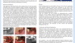
VERTICAL RIDGE AUGMENTATION BY CALLUS DISTRACTION UNDERNEATH HA-COATED TITANIUM PLATES – PROOF OF PRINCIPLE AND FIRST CLINICAL DATA
Objectives: Distraction osteogenesis is based on the separation of two vital bone segments and careful distraction after initial formation of callus. A callus between a titanium surface and surrounding bone (“jumping gap regeneration“) might also be eligible for expansion, providing a significantly less-invasive method of distraction osteogenesis. The aim of this pilot study was to evaluate the elevation of a thin HA-coated titanium plate and subsequent callus distraction beneath the titanium. Methods: After gaining approval of the local ethics committee of the University Hospital of Cologne and authorisation by the Federal Institute for Drugs and Medical Devices (BfArM, Bonn, Germany), 10 healthy patients with a vertical bone defect of the posterior maxilla or mandible (two bilateral) were consecutively enrolled. Mucoperiosteal flap elevation was performed, and HA-coated titanium plates (Bonehill Type Zä/® prototypes 14x7mm or 18x8 mm) were inserted 1–2mm away from the underlying bone, by fixing to adjacent teeth using individual bridges. Soft tissue was thickened up beneath the periosteum using a 1-mm collagen matrix (Mucodermä/®) and closed using single mattress sutures. The callus-forming phase was 7–10 days. Membranes were then elevated 0.70mm per day until the desired individual augmentation heights of 4–8 mm were reached. After 4–8 months‘ consolidation, the titanium plates were removed and bone core biopsies harvested in preparation of the implant bed. Conventional radiographs were taken at baseline, after plate insertion, and after the healing periods. Results: Initial healing was uneventful in all cases, with 8 of 12 augmented areas showing sufficient vertical bone formation at the end of the consolidation period. Implants were placed as planned, and histological evaluation showed excellent bone formation with physiological microarchitecture of the bone trephines. One site showed complete reduction of the initially achieved augmentation height based on insufficient retention; in other cases there were trauma-related soft tissue perforations with subsequent removal. In the successful cases, radiographs taken after 4-months‘ consolidation showed initial mineralisation of augmented areas, with increasing radiodensity up to 8 months. At implant uncovering, bone levels were stable radiologically and clinically in both vertical and horizontal dimensions. Conclusions: Given the limitations of this pilot study, we conclude that this novel technique of distraction osteogenesis using HA-coated titanium plates in distance to the underlying bone leads to predictable vertical bone regeneration; this is a proof of concept. It also offers a minimally invasive alternative for vertical ridge augmentation in severely resorbed jaws. A trial based on parallel inclusion of a higher number of patients in a multicentre setting using a device with an internal distraction mechanism, has now begun yielding data on new applications. -

HYDROXYAPATITE-COATED TITANIUM SUBSTRATE— INVESTIGATION OF CYTOTOXICITY
Objectives: The cytotoxicity of the hydroxyapatite (HA) coating was investigated in vitro on murine fibroblast L929 and human liver carcinoma HepG2 cell lines. Methods: The titanium substrate surface was prepared by alkali etching with sodium hydroxide, subsequent replacement of Na+ with Ca2+ ions and thermal treatment at 600°C. Prepared specimens were used as a substrate for plasma jet deposition of HA. The plasma installation PJ-100TM/® was used for the plasma spray process. HA-coated samples and the titanium substrate underwent cytotoxic analysis. The cytotoxicity of both samples was tested in vitro on a single-layer cell culture by the direct contact method according to ISO-10993-5. For examination of the cytotoxic activity of both samples, L929 (murine fibroblast) and HepG2 (human liver carcinoma) cell lines were used. Experiments were performed with cells of viability of greater than 95% and cell confluence in a culture of 80%. The cytotoxicity investigation assessed metabolic activity using 3-[4,5 dimethyl-thiazol-2-lyl]- diphenyl tetrazolium bromide (MTT proliferative activity was measured by [3H]-thymidine incorporation; necrosis by lactate dehydrogenase (LDH) and propidium iodide (PI) assays; oxidative stress by nitrite measurement; and reactive oxygen species (ROS) by flow cytometry. Cell growth intensity and morphology were evaluated by phase contrast microscopy. Results: Adhesive strength of the HA coating was 60.5± 5.5MPa. X-ray diffraction showed mainly the presence of crystalline HA. Scanning electron microscopy revealed a very rough surface of the coating with micro and nano patterns. L929 cells were more sensitive than HepG2 cells in metabolic response to analysed samples. A statistically significant reduction of 20% was observed in metabolic activity for L929 cells cultured in the presence of both analysed samples compared to untreated cells. The samples showed different proliferative activity of L929 and HepG2 cells compared with untreated cells – L929 cell proliferation was significantly higher (15–20%) and HepG2 cell proliferation was significantly lower (20%). Morphological analysis of both cells lines showed no cytopathic effect of the tested samples in the immediate and distant areas of the sample in cell culture plate wells. After removing the samples, impaired confluence of cell growth on the plate surface was found beneath the sample, but reactivity was considered non-cytotoxic due to the limited area. LDH levels indicated the absence of (or very low) cytopathic effect of the analysed samples. Low cytotoxicity was confirmed by PI staining. There was no significant change in nitrite accumulation. A significantly higher production of ROS was observed in cultures of both cell lines in the presence of HA-coated samples. Conclusions: The changes in metabolic and proliferative activity of the cells in culture in the presence of HA samples were accompanied by an almost complete absence of cytopathy; metabolic activity and parameters of cell necrosis were significantly below 30% and the coating can be considered as non-cytotoxic, and therefor promising for biological applications. In terms of ROS production, HA modification of the titanium substrate increased the oxidative response of the tested cells, also without any cytopathic implications. The biological effects of HA coatings need to be further evaluated in vivo.

Take this survey to help us improve your experience
on Dental Campus
For completion, you get 3 days unlimited access to:
- All lectures
- All cases
- All videos