


-


A STANDARD INDEPENDENT BONE REGENERATION MODEL FOR TISSUE ENGINEERING AND OSSEOINTEGRATION
Objectives: Functional tooth replacement and bone regeneration are a routine part of daily practice in modern dentistry, but there is no reproducible and relatively inexpensive animal model for multiple testing. We aimed to develop a new model for evaluating bone regeneration, osseointegration and critical size defects. Methods: We used a special guided approach at the level of the C4–C5 vertebrae in Wistar rats after cutting the tail for parallel placement of customised titanium implants. Bone remodelling around the implants was evaluated after 4–16 weeks. Biomechanical properties were characterised by measuring the maximum extraction force and using micro-CT, histomorphometry and resonance frequency analysis. Critical size defects for bone tissue engineering and implant placements for titanium body experiments were evoked by drilling each of the first four caudal vertebrae from the side, thus allowing multiple comparisons, and micro-CT and histology were used to evaluate bone growth. Results: The percentage regeneration was only of 20% ± 5% when bone grafting particles alone were used to fill defects. Bio-Oss[tm] was used for positive controls but the material was radio-opaque and could not be assessed by micro-CT. Histology revealed collagen network formation in defects filled with bone grafting material, but not in those with Bio-Oss[tm]. Immunohistochemical assays confirmed the presence of bone markers such as osteocalcin and type I collagen, and the presence of bone cells of both human and rat origin. A positive correlation was found between measurements made using various in vivo techniques(r=0.8498). Statistically significant increases were found during evaluations at 4, 8, 12 and 16 weeks in terms of strength of osseointegration. Histomorphometry and micro-CT showed new bone formation on the implant surfaces, suggesting the direct connection occurring between bone and titanium. Conclusion: These results support the use of the caudal vertebrae bone regeneration rat model as a useful standard for preclinical evaluation of tissue engineering and osseointegration. -
AN OLD-SCHOOL SOLUTION TO A NEW-AGE PROBLEM—RIDGE MAPPING FOR IMPLANT PLANNING
Objectives: Understanding the bony anatomy at the site of implant placement is essential for implant success. Computed tomography (CT) is generally considered the gold standard for assessing bone morphology, however there are situations in which it is not practical. The ridge-mapping technique has been advocated as a convenient way to assess site suitability for implant placement, and was used in this patient to plan a single dental implant. Methods: An unrestorable maxillary lateral incisor tooth was extracted and the socket was preserved using Bio-Oss[tm] and a Bio-Gide[tm] membrane. This healed undisturbed for 2 months, then alginate impressions were taken and plaster study models fabricated. A mapping stent was fabricated from the plaster model using rigid acrylic material to ensure stabilisation[stability?]. The stent overlapped the buccal and palatal mucosal tissue. Holes were drilled in the stent at 2mm, 4mm, 6mm and 8mm from the alveolar crest, allowing a size 20 endodontic file to pass through. At each pre-drilled site, the depth was measured from the acrylic stent surface to the bone. The cast was then sectioned and measurements were transferred to the cast to provide an outline of the underlying bone. Following clinical assessment and outline measures, a bone level Strauman implant of 3.3-mm diameter and 12-mm length (SLActive was planned. Local anaesthesia was administered before making a slightly palatal incision and reflecting a full-thickness mucoperiosteal flap. The alveolar ridge was measured using a caliper. The osteotomy was prepared using standard drills and the implant was placed 3mm from the cervical margin of the proposed restoration. Results: Ridge mapping and direct measurement with calipers yielded similar dimensions of the alveolar crest. Conclusion: This report demonstrated that ridge mapping using endodontic files and a model-based template gives an accurate representation of the underlying alveolar bone. This technique may be suitable when standard radiographic techniques are not possible. -
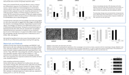
SALIVA SUPPORTS THE FORMATION OF PRO-INFLAMMATORY MACROPHAGES IN MURINE BONE MARROW CULTURES
Objectives: The process of oral wound healing requires a temporary inflammatory reaction. Saliva can support this process, as reported previously, by provoking a strong inflammatory response in oral fibroblasts, while suppressing osteoclastogenesis in bone marrow cultures. Bone marrow cells also give rise to a heterogeneous cell population of macrophages which are also a involved in wound healing. Lipopolysaccharide (LPS) and interleukin(IL)-4 both induce activation of pro-inflammatory and anti-inflammatory macrophages (M1 and M2, respectively), but he impact of saliva on programming bone marrow cells into either M1 or M2 macrophages remains unclear. Methods: Fresh sterile saliva, similar to LPS, was used in culture of mouse bone marrow to observe its affect on the process of macrophage polarisation. Evaluation was conducted using real-time gene expression analysis and immunoassays for various indicators of different macrophage types. Results: Fresh sterile saliva was associated with robust activation of the M1 phenotype, characterised by a strong increase in the genes for interleukins IL-12, IL-6 and Nitric oxide synthase-2 (NOS2) observed with real-time gene expression analysis and immunoassay. It was also associated with increased expression of IL-10, which is characteristic of the M2 phenotype, but not the proteins Ym1, FIZZ-1 and arginase. Conclusion: These preliminary in vitro results show that saliva can support the formation of pro-inflammatory macrophages. We are currently evaluating the role of TLR4 signaling with the antagonist TAK-242, and the respective downstream signaling pathways, as well as the impact of the saliva pellicle on M1 or M2 macrophage activation.

Take this survey to help us improve your experience
on Dental Campus
For completion, you get 3 days unlimited access to:
- All lectures
- All cases
- All videos