


-

RIDGE PRESERVATION OF EXTRACTION SOCKETS WITH CHRONIC PATHOLOGY USING BIOOSS COLLAGEN WITH OR WITHOUT BIOGIDE—AN EXPERIMENTAL STUDY IN DOGS
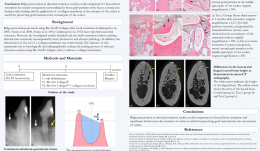
Objectives: Ridge preservation protocols using BioOss Collagen after tooth extraction have successful outcomes, but previous studies included only fresh extraction sockets and omitted infected sites with chronic pathology. The effectiveness of collagen membranes is also controversial. The aim of this study was to histologically and radiographically evaluate the healing process of infected extraction sockets using BioOss[tm] Collagen with or without collagen membrane. Methods: Six adult Beagle dogs aged 1–2 years, and approximate weight 10 kg, were used. Sulcular incisions were made, and a flap was elevated in the mandibular premolar area. The left third mandibular premolar and both fourth mandibular premolars were hemisected with fissure burs, and the distal roots were removed atraumatically. A #2 carbide round bur was used to dig a notch in the mesial side of the mesial root, and the notch was filled with a collagen sponge soaked in Porphyromonas gingivalis. The pulp of the mesial roots was removed with a K-file before injection of P. gingivalis and sealed with intermediate restorative materials before suturing the flap. Following communication of the two pathologies (after about 4 months), the mesial roots were extracted and the defects were randomly assigned to one of three subgroups using a 3 x 3 Latin square design. These were no treatment (controls); BioOss Collagen graft (group T1); and BioOss Collagen graft with BioGide collagen membrane (group T2). After 7 months from induction of the lesion (baseline), the dogs were sacrificed and block biopsies were placed in a fixative, dehydrated and embedded. The percentage of different tissues (mineralised bone, bone marrow and biomaterial) in the infected extraction sockets was evaluated, and each extraction site specimen underwent the micro-CT examination. Results: Micro-CT revealed differences in height between the buccal and lingual crests, in addition to the bone morphometric parameters. There were significant differences in the vertical distance between buccal and lingual crests in controls (2.22 mm ± 0.26) and T2 (1.80 mm ± 0.16. Bone surface density (BS/BV) values were 0.32 ± 0.05 (controls), 0.45 ± 0.13 (T1) and 0.50 ± 0.03 (T2). There was a significant difference between BS/BV values in controls and T2. The level of interconnectivity of the bone (TbPf) gave values of –0.53 ± 0.24 (controls), –0.44 ± 0.11 (T1) and –0.32 ± 0.15 (T2). Values for structural model index (SMI) were –5.34 ± 3.07 (controls), –2.94 ± 2.38 (T1) and –1.27 ± 0.83 (T2). TbPf appeared to increase gradually from controls to T1 then T2. The sockets in the control group were occupied by mineralised bone (68.08% ± 6.51) and bone marrow (30.56% ± 6.07%); in T1 they were filled by mineralised bone (55.90% ± 4.70%), bone marrow (23.50% ± 4.19%) and BioOss[tm] particles (18.49% ± 2.11%), and T2 contained mineralised bone (61.38 ± 6.9%), bone marrow (10.31% ± 4.93%) and BioOss[tm] particles (27.04% ± 5.25%). The T2 sockets contained more bone graft material than those in T1. There was a significant difference between amount of mineralised bone in controls and T1. Conclusion: In the micro-CT analysis, the vertical distance between the lingual and buccal crests was significantly shorter in the T2 group (ridge preservation with BioOss Collagen and collagen membrane) compared to controls. This is in line with a previous study that reported similar results using BioOss Collagen for ridge preservation, indicating a possible role of the biomaterial in promoting de novo bone formation to maintain the ridge profile and compensate for marginal ridge resorption. The use of a membrane for ridge preservation (as in T2) to prevent loss of the graft material in the initial healing phase may have improved contact osteogenesis near the coronal part of the buccal bone. Ridge preservation in infected extraction sockets may compensate for buccal bone resorption, and using membrane to cover the entrance of sockets may help preserve graft material near the coronal part of the socket. -

FIRST CLINICAL EXPERIENCES WITH INDIVIDUALISED 3-D AUGMENTATION WITH CAD-CAM DESIGNED TITANIUM MESH ReOss
Objectives: The augmentation of the jaw has been, and continues to be, a sophisticated therapy in implantology, especially when vertical or combined defects have to be restored. Modern computer aided CAD-CAM technologies have led to the revival of old and established augmentation techniques, such as the use of titanium mesh for onlays and guided bone regeneration techniques. ReOss[tm] provides an individualised CAD-CAM-based titanium mesh based on CT, DVT or DICOM (digital imaging and communications in medicine) patient data. Methods: The augmentation possibilities with this new system were evaluated in a small clinical trial of patients (n = 15) with seventeen different defect regions. DVT-based DICOM (digital imaging and communications in medicine) data of patients who needed horizontal, vertical or combined augmentation were used to create an individualised titanium mesh. For the augmentation, a mixture was prepared of autologous bone mostly harvested from the lower jaw and BioOss[tm] particles (1:1) was used. All defects were covered with BioGide[tm] in combination with platelet-rich fibrin (PRF) membranes. All patients received antibiotic therapy for five days. After 6 months, re-entry was performed, with explantation of the titanium mesh and simultaneous implantation. Results: The patients (27% men and 73% women; average age 34 (range 18–60) years) underwent augmentation in the lower jaw (n = 12) and upper jaw (n = 5). Of these, 47% had a horizontal defect, and the remainder had a vertical or combined defect. Augmentation was performed to replace two to seven teeth (an average of three). In all cases, the tailormade titanium mesh was easily placed in the planned area of augmentation. Exposure of the mesh during the healing period occurred in 29%. There was no total loss of the augmentation, nor did exposure lead to situations in which planned implantation could no longer be carried out. All cases showed sufficient augmentation volume that was congruent with the preoperative planned augmented volume. Conclusion: Individualised ReOss[tm] CAD-CAM titanium mesh provides a sufficient onlay and guided bone regeneration technique for augmentation of jaws. The soft tissue covering remains one of the most critical steps in this procedure, and quick and easy application is a great advantage even in vertical defects. -
IMMEDIATE LOADING OF THE MAXILLA USING FLAPLESS SURGERY TO INSERT ONE-PIECE ZIRCONIA IMPLANTS IN PREDETERMINED POSITIONS
Objectives:In the aesthetic area, immediate implant placements are increasingly common. Using a 3-D custom-made guide for insertion is associated with less trauma and faster healing periods. Titanium implants may cause unfavourable soft tissue conditions or recession of gingiva in the area, and lead to compromised aesthetic outcomes. In this study, zirconia implants were chosen rather than titanium.. Methods:A 18-year-old woman, who had previously suffered serious trauma of frontal maxilla, was referred to our dental practice. Due to external root resorption, the central incisors were removed 15 months previously, and she had a fixed orthodontic retainer with two substitute acrylate teeth (#11 and #21) to preserve jaw line functionality and aesthetics. The patient was still adolescent, but thorough examination by cone beam CT established that her jaw development and growth was complete. We decided to insert two zirconia implants, with a comprehensive procedure because the bone loss at both positions was severe. The first step was horizontal augmentation of the alveolar ridge using a membrane technique in order to preserve necessary bone (BioResorb Macro Pore[tm]). Six months later, two zirconia implants (SDS, ATZ, 3.8x11.0mm, torque 35N/cm) were inserted using flapless surgery into predetermined positions using a 3-D custom-made guide. Two connected provisory acrylate crowns were inserted immediately, to stabilise the implants and give the patient functional teeth for the following five-month period. The crowns also aided the process of implant osteointegration, providing an ideal emergence profile and supporting the peri-implant soft tissue. Follow-up was uneventful. Three months after surgery, the final prosthetic restoration comprised of two connected ceramic, metal-free crowns. Results: The healing period was uneventful with no adversary reactions. Both procedures resulted in minimal trauma, due to the use of 3-D custom-made guides for both bone regeneration and implant insertion, which led to a good post-operative period. The first procedure (bone regeneration) gave satisfactory results, and the second (implant placement) gave excellent primary stability without radiographic bone loss or gingiva inflammation. Peri-implant soft tissue parameters showed promising results. Probing depth, clinical attachment level and modified bleeding index at the implant sites were all satisfactory. The restoration work showed a healthy gingival margin and no discoloration of the soft tissue. The most important patient feedback related to permanently improved perception of function, aesthetics, sense, speech and self-esteem (which is very important in adolescents). Conclusion: Use of 3-D custom-made guides for implant placement enables faster and better recovery because trauma is minimised. There is an increasing demand for metal-free implants because of the potential immunologic and aesthetic compromises associated with titanium implants entails. Unfavourable soft tissue conditions or gingival recession may lead to compromised aesthetics, which are particularly concerning when maxillary incisors are involved. Cervical bone loss and gingival recession associated with metal implants tend to cause bluish discoloration of the overlying gingiva, thus impairing aesthetic outcome. Zirconia implants are a good choice for the aesthetic zone. In our case, the gingival margin was healthy and there was no discoloration of the soft tissue. Zirconia implants are also suitable because they have a tooth-like colour, good mechanical properties (high strength, fracture toughness) and good biocompatability. -

ADVANTAGES AND DISADVANTAGES OF COMPUTER-DESIGNED TECHNIQUES FOR ORAL REHABILITATION - A REVIEW
Objectives: In the last four decades, dental implants have become an essential part of oral rehabilitation. Successful implant placement depends directly on highly accurate treatment planning and surgical procedures. It is important to review the accuracy data for surgical and prosthodontical techniques such as guided surgery. Methods: A retrospective review was conducted for cases using an image-guidance system for the accurate placement of implants. An electronic literature search of PubMed database was conducted, combining controlled terms and keywords whenever possible. The search terms were “computer-guided design”, “image-guided surgery”, “computer-assisted manufacture”, “computed tomography” “dental implants” and “surgical guide”. Articles from January 2005 to September 2015 were screened. They were included in a qualitative assessment if they were recent papers and had references to articles related to the terms defined above. Results: Software guides may be useful for decreasing the incidence of implant-associated difficulties, as well as achieving greater precision in placement of implants, leading to better preservation of anatomic structures (especially for full-mouth rehabilitation). The most common difficulties encountered during placement are improper location, wrong angulation, and lack of primary stability. These techniques provide sufficient information about the bone anatomy to avoid these issues and the treatment plan reduces operating time and surgical trauma and increases implant success. However, these techniques require knowledge of and experience in the use of 3-D information for virtual planning and implant positioning, and there is a lack of visibility and tactile control during the procedure, thus vital anatomical structures can be damaged. The available literature on computer-aided surgical guidance has only limited data and relatively short observation periods, therefore further research should focus on clinical studies with long-term follow-up. Conclusion: Surgical implant complications are not uncommon and should be addressed immediately. Causality may be iatrogenic due to poor treatment techniques, and a successful final treatment can be possible by the use of computer-aided surgical guidance. Competent surgeons yield acceptable to excellent results and surgery is predictable using these techniques, but care should be taken when using them. -

APPLICATION OF BIO-OSS WITH FREE GINGIVA GRAFT FOR MAINTAINING SOCKET CONTOUR IN IMMEDIATE IMPLANT PLACEMENT
Objectives: To evaluate the application of BioOss with free gingival graft for maintaining the socket contour in immediate placement of implants. Methods: After extraction of a fractured 21# tooth in a 50-year-old male patient, with thick gingival biotype, a flapless immediate placement implant (4.1 mm x 12mm Straumänn SLA was inserted to the tissue level of a standard implant, and covered with an insertion screw. Grafting was with 0.25g Bio-Oss small particles, using free gingiva graft from palatal epithelium to close the socket. The natural tooth was cemented with a crown for immediate restoration . After 3 months, the second-stage soft tissue was shaped with provisional restoration by temporary abutment and the natural crown. After another 6 weeks, impressions were taken and the final PFM (porcelain-fused-to-metal) crown was fixed with screw retention. Results: Within 4 years, the soft and hard tissue levels around the implant were stable, and the crown was supported by the implant with a natural profile with the neighbouring teeth. Conclusion: Immediate implant placement covered with BioOss and free gingiva graft can result in a long-term stable aesthetic outcome in patients with thick gingiva sites. -
EFFECT OF A 0.2 % HYALURONAN-CONTAINING GEL ON ALVEOLAR BONE REMODELLING IN RATS WITH EXPERIMENTAL PERIODONTITIS
Objectives: Hyaluronic acid (HA) is a naturally occurring linear polysaccharide of the extracellular matrix of connective tissue and other tissues. It has several physiological and structural functions, accelerating bone regeneration by means of chemotaxis, proliferation and successive differentiation of mesenchymal cells. The purpose of this study was to evaluate the effect of topical application of a 0.2% hyaluronan-containing gel on bone remodelling in the mandible of rats. Methods: Thirty-two white Wistar rats (weighing 350–450 g and aged 4 months) were divided into three groups: group 1 comprised intact rats (control); group 2 comprised rats with a “peroxide” model of periodontitis, reproduced by a 60-day diet of over-oxidised sunflower oil (model); and G3 comprised rats with peroxide-induced periodontitis that were given topical HA-gel on the gingiva daily for 14 days (treatment). Alveolar bone remodelling was evaluated by biochemical and histologic analysis, and activity of alkaline phosphatase (ALP; an indicator of osteoblastic activity) and tartrate-resistant acid phosphatase (TRAP; a biomarker of osteoclasts) were measured in the alveolar bone. Results: The enzyme activity in alveolar bone was significantly higher in the treatment group than the model group, with levels of 42.56 ± 3.96 mckat/kg and 79.94 ± 3.17 mckat/kg (p Conclusion: The results suggest that topical application of a 0.2% HA-containing gel applied to the gingiva in rats has an osteoinductive effect. -
MODELLING CRITICAL-SIZED BONE DEFECTS IN RABBIT MANDIBLE: A MORPHOLOGICAL STUDY OF BONE HEALING
Objectives: There is little consistency in the choice of appropriate animal models in bone research, where evaluating bone repair dynamics is a basic part of bone morphofunctional studies. Among the available models, rabbits have several advantages. The bone defect repair rate is mainly dependent on the size of the bone wound, and experimentally created osseous injuries for studying repair mechanisms should be wide enough to preclude spontaneous healing. This study aimed to assess bone healing histologically in rabbit mandible defects. Methods: Three adult male rabbits were used. Defects with diameters of 3mm and 6mm, and a depth of 1.5mm, were created by excision of the mandible cortex using a trephine. Defects were allowed to fill with blood and the overlying soft tissues were repaired. Postoperatively, the rabbits were given antibiotic and analgesic therapy. In order to assess osteogenic activity, vital fluorochromic bone markers were used. After 6 weeks surgery, the animals were euthanised and bone segments were dissected and processed for histological evaluation. Analysis was performed with a light microscope. Results: During the observation period, the animals were healthy and presented no signs of infection. Both sagittal and transverse sections showed the dynamic of injury healing. After 6 weeks, the 3-mm defect had completely healed without inflammation, with proliferation of fibrous connective tissue and a minimal amount of immature woven bone. The new bone contained numerous blood vessels and osteoblasts. The 6-mm defect did not fully heal. Bone repair tissue was characterised by woven fibres, consistent with immature primary bone, and primitive Haversian spaces. Conclusion: We investigated the kinetics of healing in different sizes of rabbit mandibular defects. The 6-mm defect was the critical-sized defect. This experimental model will allow evaluation of the effect of different biomaterials on the healing process, in terms of adequacy of tissue quantity and quality, as well as the osteoconductive and osteoinductive characteristics of the test materials. -
ROOT APPROXIMATION AND PENETRATION OF MAXILLARY SINUS AND ITS INFLUENCE ON POST-EXTRACTION OROANTRAL COMMUNICATION— A NOVEL TECHNIQUE FOR OROANTRAL COMMUNICATION REPAIR, AUGMENTATION, AND FINAL RESTORATION
Objectives: The objective of this study was to evaluate the approximation of maxillary tooth roots to the sinus, the incidence of root penetration into the sinus, and its influence on extraction of maxillary posterior teeth. A technique for repairing oroantral communication aimed to augment the extraction site and provide valuable bone volume for implant insertion and final restoration. Methods: Forty-six patients were evaluated by x-ray and perpendicular measurement of sinus approximation from the tip of the root of the posterior teeth to the sinus. The number of root penetration into the sinus were counted. Ten cases had oroantral communications post-extraction and were enrolled in the study. The repair intervention was delayed in two, and eight were operated on at the time of extraction. Sinus floor was elevated noninvasively through the socket and collagen matrix was inserted into the lateral wall of the sinus and rotated to close the oroantral communication of the palatal wall. Corticocancellous bone chips were used to augment the site. Collagen matrix was used again to cover the site, with soft tissue closure. The sites were evaluated by x-ray four months later, to evaluate bone volume and density. Implants were inserted and final restoration was done after four months. The average follow-up of patients as 2.5 years. Results: Both the left and right sides were evaluated for sinus approximation and root penetration. The combined average distances were recorded for the upper first premolars (9.4mm), upper second premolars (4.55mm), upper first molars (4.065mm), upper second molars (1.87mm) and upper third molars (3.6mm). The mean percentage penetration was calculated for the upper first premolars (1.25%), upper second premolars (8.53%), upper first molars (46.4%), upper second molars (27.65%) and upper third molars (21.82%). In this case series, all oroantral communications were successfully closed and all sites were augmented without postoperative complications. Clinical and radiographic evaluations revealed successful implant restoration. Conclusion: Within the limitations of this study, oroantral communications can be repaired and augmented for implant restoration by proper preoperative evaluation of sinus approximation using this novel technique. -
PERI-IMPLANT SOFT TISSUE STABILITY AFTER SINGLE IMPLANT RESTORATION USING EITHER GUIDED BONE REGENERATION OR A CONNECTIVE TISSUE GRAFT . A RANDOMIZED CLINICAL TRIAL
Objectives: The primary purpose was to compare two different surgical procedures: the connective tissue graft and the GBR technique, in conjunction with implant placement. A secondary aim was to assess the influence of the two different procedures on periodontal soft tissue stability of teeth adjacent to the implant site. The null hypothesis was that connective tissue grafting procedures can produce similar clinical results to GBR. Methods: A total of 32 patients were included in the study. Prosthetic stage was scheduled after 6 months from implant insertion. Results: Between-group (test versus control) differences in buccal mucosa thickness were not statistically significant (p = 0. 669). Both groups showed statistically significant differences in changes over time (p = 0.023), with a statistically significant increase in mucosal thickness between T0 and T1 (p = 0.021). There were statistically significant intra-group differences in keratinised tissue height at the implant sites (p Conclusion: The one-year results of this clinical trial on 32 consecutively treated patients confirmed previously published data at 1 and 3 years. The measured clinical parameters indicated healthy and stable peri-implant soft tissue over time in both the test and control groups. Similar results were found for adjacent teeth, thus it may be argued that the coronal reposition of the envelope flap maintains the stability of periodontal tissues following both bone and connective tissue grafting procedures. Within the limitations of this study, it can be concluded that the connective tissue graft procedure was beneficial in maintaining facial gingival level when performed in conjunction with implant placement, showing no statistically significant differences in term of mucosal shrinkage with GBR. -
HISTOLOGICAL ANALYSIS OF CORES OBTAINED DURING IMPLANT PLACEMENT IN SITES WITH PREVIOUS LATERAL MAXILLARY SINUS FLOOR LIFT
Objectives: Recent studies using PCR and RT -PCR analysis has verified that the internal structures of implants and abutmant are heavily contaminated with pathogenic bacteria. The purpose of this study and topographic characterization of the internal spaces (Endoimplanto) of the implants using 2D and 3D computed microtomography. Methods: Six implants Biomet External hexagon cylindrical implants with tapered apex (3.75.0 x 11.5 mm Pi-Brånemark, EXOPRO, Bauru, Brazil) were used to test. Inner structure of implants and abutmant was divided into 4 zones: Endoimplanto/UCLA, Endoimplanto/Abutment, Endoimplanto/Microgap, EndoImplanto/Implants. All micro-CT scans were acquired on a Triumph multi-modality system (Gamma Medica, Northridge, CA) using the following acquisition parameters: tube voltage 80 kVp, tube current 250 A, detector pixel size 50 m, focal spot size 50 m and a field-of-view of 59 or 93 mm. The spatial resolution was 39 m and 28 m, and the radiation dose was 8.3 cGy and 19.7 cGy for a field-of-view of 93 mm and 59 mm, respectively. Results: The results showed that the endoimplanto abutment/UCLA corresponds to 40/50%, Endoimplanto/Abutment 10-20% , Endoimplanto/Microgap 2-3 %, Endoimplanto/Implant 10 -15 % of full structucture implant/Abutment /UCLA. Conclusion: It concludes that the empty spaces of the implants (Endoimplanto ) occupy a large part of the overall volume Implant /Abutment /UCLA. This space can serve as a repository of relevant bacteria and fungi in the development of periodontal and peri-implant disease. The microtomography and a good non -destructive and non-invasive method to search the internal structures of the implants. -
PERIOSTEAL PREFABRICATION OF VASCULARISED CAD/CAM BONE SCAFFOLDS
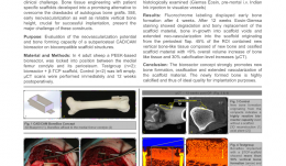
Objectives: Reconstruction and prosthetic rehabilitation of patients presenting with severely atrophied alveolar ridges or critical-size bone defects of the facial skeleton are clinically challenging. Bone tissue engineering with patient-specific scaffolds are a promising alternative for overcoming the drawbacks of autologous bone grafts. Early neovascularisation and reliable vertical bone height are both crucial for successful implantation, and present the major challenge of these constructs. The aim of this study was to evaluate the neovascularisation potential and bone-forming capacity of a CAD/CAM-designed artificial space (bioreactor) between the medial femur condyle and its periosteum on bioresorbable scaffold structures. Methods: Four skeletally mature Swiss alpine sheep were used. The bioreactor, composed of a PEEK construct (side walls), a vascular pedicle flap (roof) and deperiosted compacta (floor), was locked into position by two 2.4 standard screws. The sheep were put in two groups of two: in the treatment group, the created rectangular space was filled with a bioresorbable β-tricalcium phosphate (β-TCP) scaffold; in the other group, the bioreactor was left empty (negative control). Micro-CT scans were performed immediately and 12 weeks postoperatively. Fluorochrome labelling was performed with calcein green after 4 weeks, and with Xylenol orange after 8 weeks to document new bone formation. Animals were euthanised 12 weeks postoperatively and surgery sites were examined histologically using eosin–Giemsa with pre-mortal intravascular injection of Indian Ink to visualise blood vessels. Results: Fluorochrome labelling displayed moderate early bone formation after 4 weeks with already in-growth into the bioreactor. After 8 weeks, there was pronounced new bone formation in the bioreactor. After 12 weeks, eosin–Giemsa staining showed degradation and bony replacement of the scaffold material, bone in-growth into the scaffold voids, and extended neo-vascularisation into the scaffold originating from the periosteal flap. Micro-CT data showed that after 12 weeks of implantation, 45% of the region of interest (ROI) contained new vertical bone-like tissue composed of new bone and ossified scaffold material. The overall volume increase of bone-like tissue in the ROI was +9% and the calcification level increased by 30%. Conclusion: These results indicate that the bioreactor concept strongly promotes new bone formation, ossification and extended vascularisation of the scaffold material. The newly formed bone is highly calcified and thus of ideal quality for implantation purposes. -
VOLUMETRIC SOFT-TISSUE CHANGES WITH THREE DIFFERENT RIDGE AUGMENTATION PROCEDURES—AN EXPERIMENTAL STUDY IN BEAGLE DOGS
Objectives: Guided bone regeneration by means of membranes and bone substitutes is one of the most documented approaches for restoring deficient alveolar ridges. Despite routine use of the techniques, it is not known to what extent these approaches can restore the tissue volume lost after tooth extraction. The aim of this study was to analyse the volume loss occurring after extraction and how much lost volume can be recovered by three different bone augmentation procedures. Methods: To create the alveolar ridge defects, the teeth were sectioned in a buccal–lingual direction at the furcation level, and the mesial roots of M1 and P4 and distal roots of P3, P2 and P1 (monoradicular) were individually extracted. Three vertical grooves of about 6mm in height and 6mm in depth (as far as the lingual cortical bone) were prepared in order to eliminate the buccal bone plate. After 12 weeks, the dogs were randomly assigned to one of three regenerative procedures: graft-only (B) in which the defect was filled with a particulate bone substitute (Bio-Oss Collagen); membrane only (M) in which the defect was covered with a bioabsorbable collagen membrane (Bio-Gide); and combination group (T) in which the defect was filled with a particulate bone substitute (Bio-Oss Collagen) and membrane was added. Conventional polyvinyl impressions were taken before extraction and defect creation at one time point (T1), before regenerative surgery (T2) and after 3 months of healing (T3). The cast models were optically scanned to obtain stereolithography (STL) files. Linear and volumetric measurements included volumetric analysis, distance between surfaces, and tissue thickness at three levels below the alveolar ridge on the buccal side of the regenerated site(at 1, 3 and 5mm). Results: A total of 18 casts were obtained at T1, with 18 at T2 and 9 at T3. No impressions were taken at the time of sacrifice to avoid disturbing early healing. The box-shaped defects were effective in creating horizontal and vertical ridge deformities (comparison T1–T2). None of the three regenerative approaches resulted in complete reconstitution of the alveolar ridge to baseline dimensions (comparison T1–T3), although volume gain in groups B and T was significantly better than in groupM (comparison T2–T3). Conclusion: The three regenerative approaches failed to rebuild the post-extraction loss in tissue volume.

Take this survey to help us improve your experience
on Dental Campus
For completion, you get 3 days unlimited access to:
- All lectures
- All cases
- All videos