


-

ALVEOLAR RIDGE RECONSTRUCTION USING A SPLIT-CREST TECHNIQUE IN ATROPHIC MAXILLA WITH BIO-OSS AND BIO-GIDE —TWO CASE REPORTS
Objectives: In clinical cases where a severe horizontal bone deficiency is present, it is very difficult to install implants in the perfect position for achieving optimum aesthetics and functional rehabilitation. The split-crest technique leads to reduced waiting times for installation of implants for maxillary defects of less than 3mm. The cases reported here underwent the split-crest technique with interposition of Bio-Oss biomaterial and were reopened for implant placement after five months. Methods: Patients were selected with a horizontal bone deficiency in the maxilla of 1.6–2.3mm. Both were fit for surgery. The preoperative assessment consisted of blood tests, CT scans and antibiotic prophylaxis and full treatment plans were presented to the patients. Infiltrative terminal anesthesia was applied until satisfactory tissue ischaemia was produced. An incision was made above the crystal with 15c blades and mesiodistal relaxing incisions. After total detachment and bone mucoperiostal exposure, a tag 701 was made with a surgical drill at the cortical level, completed with a metal disc osteotomy. A chisel was used between the markings to achieve total displacement of cortical , to achieve sufficient expansion and an intact nasal spine . Screws were placed at the base of each fracture, preventing vestibular cortical blades from moving during bone maturation. Gaps and remaining spaces were filled with Bio-Oss. Bio-Gide membranes were applied, the flap repositioned and the wound was closed by suturing without tension. Results: CT scans four months postoperatively showed increased bone width of approximately 5.2mm. There were no complications and both patients reported little discomfort after surgery. Both underwent further surgery for implant placement after five months. Primary stability of all implants was achieved and manufacturing the prostheses. Conclusion: The split-crest technique is a feasible option. Using Bio-Oss with a Bio-Gide membrane increased the maxillary bone thickness, which facilitated rehabilitation with dental implants. -
IMMEDIATE LOADING WITH ONE-SHOT PROSTHESIS (OSP) AND RECURRENT IMPRESSION (RI) FOR EDENTULOUS PATIENTS TREATMENT—A CASE SERIES
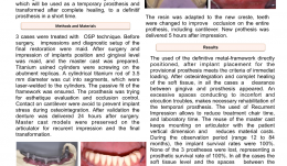
Objectives: Immediate-load protocols for edentulous patients are well-documented treatment options, however it is expensive to fabricate a provisional prosthesis that is capable of loading, which must be followed after osteointegration and soft tissue healing by placement of a definitive restoration that fills gaps that appear between the gingiva and prosthesis. The aim of this study was to carry out the OSP technique using a single prosthesis with a metal framework that permits immediate loading, which could be transformed after complete healing to a definitive prosthesis. Methods: Three patients were treated by the OSP technique. Impressions were taken before surgery, with diagnostic set-up for the final restoration. After surgery, further impressions were made of the implants in position and gingival levels, and the master cast was prepared. Titanium cylinders were screwed onto the abutment replicas, and sections of 3.5-mm diameter titanium rods were laser-welded to them. Passive fit of the framework was checked. The prosthesis was intended to achieve optimum aesthetics and occlusion control. Contacts with the cantilever were avoided to prevent implant stress during osteointegration. After validation, the dentures were placed 24 hours after surgery. Master cast models were preserved on the articulator. After osteointegration, gaps between gingiva and prosthesis were recorded by taking mucostatic RIs of the edentulous areas between implants, with injection of silicone directly under the screwed prostheses. Master models were modified by eliminating the stone between the implants, and transitional silicone prostheses were screwed into place. New plaster was poured into the space to modify the crest profile according to the new anatomy, and resin was applied to improve occlusion of the teeth with the entire prosthesis, including the cantilever. The new prostheses were delivered five hours after making impressions. Results: During the observation period of 12–84 months, implant survival rates were 100%. None of the three prostheses were lost, thus the prosthetic survival rate was also 100%. In all three cases, soft tissue levels and the spaces between the prostheses and tissues were stable. Conclusion: The cost of implant treatment is important to patients. The OSP technique with RIs allows a single prosthesis to be used as temporary prosthesis suitable for immediate loading, that can be transformed, after a short time, to a definitive prosthesis that is adapted to the tissue after complete healing. This leads to cost reductions and increased access of patients. The use of RIs decreases chair time and laboratory time, and re-use of the master cast decreases material costs. Further studies are now required with larger sample sizes to better evaluate this promising low-cost option. -
PHOTOELASTIC ANALYSIS OF OBLIQUE FORCES ON DIFFERENT IMPLANT DIAMETERS
Objectives: The study aimed to review, using photoelastic analysis, the stresses generated on narrow-diameter and regular-diameter implants by simulated masticatory loads, to enable safe use of implants with different diameters. Methods: The photoelastic models were dipped in mineral oil in a polariscope. A digital NIKON COOLPIXTM 4500 camera (with lens 1:2. 6–32mm) was used to visualise the fringes and record digital images. Initial models were obtained from the silicone matrix laboratory, with a space of 10x 40mm (45mm deep) filled with plaster type 4. Photoelastic models were made by drilling the plaster with a high-speed drill and installing the analogues (standardised using a parallelometer). We planned the ideal location of narrow- and regular-diameter implants (2.8mm and 4.0mm, respectively) in the region of maxillary lateral incisors, canines and premolars. Models used in similar positions, were moulded with transfer dyes of aid to open tray. After moulding, the new analogues were screwed into resin. Crowns manufactured from titanium were via CAD-CAM using a Cerec technique were cemented onto the implants to allow better analysis of the stresses around them. Results: Loads of up to 160N were applied obliquely to the titanium laterals of the incisor crowns. Caraga of up to 250 N were placed axially to the crowns of canines and bicuspids. Conclusion: Coherent planning of rehabilitation to reduce toothless regions is important for improving predictability when using implants with reduced diameters, and to enable a lower transfer of force to the structures involved, such as the adjacent bone. It is hoped that these results will enable narrow-diameter implants to be used more safely. Finite element analysis studies do not represent the clinical reality, but all the above must be taken into consideration when planning treatment of edentulous patients. -
WELDING OF PERIODONTAL TISSUE WITH LASER-ACTIVATED INDOCYANINE GREEN (ICG) MEMBRANE
Objectives: Surgical treatment of periodontal disease requires tight closure of the surgical wound, but suturing carries the risks of infection, wound opening and scarring. Chitosan membranes with added indocyanine green (ICG) offer an alternative to suture materials and barrier membranes. The aim of the study was to assess the usefulness of ICG–chitosan membrane for the closure of surgical wounds and as a substitute for membranes for guided tissue regeneration. Methods: In this in vitro experiment, we studied 50 extracted human teeth, 30 samples of porcine gingiva and 10 samples of porcine oral mucosa. We produced chitosan–ICG membranes of 5 x 7mm and welded them with a diode laser to the gingiva, oral mucosa and surface of the tooth roots. Histological analysis was used to identify thermal damage. A rise in temperature on the surface of each sample during welding was determined from temperature profiles recorded with ThermaCAM P45TM thermal camera. The strength of linkages of the ICG membrane with the tissues were measured with a universal tearing machine (Instron 4301TM). We used parametric t-tests, ANOVA and the least significant difference (LSD) post-hoc test to analyse mean maximum temperatures and forces required to rupture linkages in the different tissue types (using s statistical significancelevel of Results: Laser welding of chitosan–ICG membrane to the gingiva, mucosa and root surface was successful and no samples showed significant thermal damage The maximum temperature measured on the root surface during welding was 42.6 ± 9.5° C, which was significantly lower than that for the gums at the already welded membrane on the tooth (55.2 ± 8.0° C) (p = 0.005). Tear forces in three groups of laser-welded gingiva and oral mucosa were 0.14 ± 0.05 N for gingival epithelial welding, 0.06 ± 0.01 N for gingiva welding to connective tissue, and 0.06 ± 0.02 N for oral mucosa welding. Tear forces in four groups of membranes laser-welded to the root surface were 1.41 ± 0.14 N for samples at an angle of 0°, 0.89 ± 0.15 N at 30°, 0.80 ± 0.14 N at 60° and 0.52 ± 0.16 N at 90°. It was 0.85 ± 0.18 N for gingiva welded to the root surface. Conclusion: Within the limits of this study, laser welding of oral mucosa and gingiva can complement the closure of mucosal and periodontal wounds, but cannot replace suturing. The favourable tensile strength of chitosan–ICG membrane linkages with root surface and chitosan’s properties in the membrane is promising as a replacement of non-resorbable membranes. Laser welding of gingiva to the root surface may add further stability to closure of the wound. -
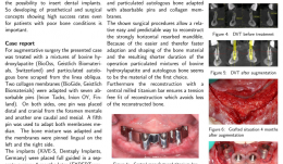
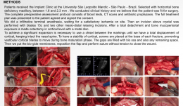
SURGICAL AND PROSTHETIC RESTORATION OF HORIZONTAL RESORBED EDENTULOUS MANDIBLE—A CASE REPORT
Objectives: Few treatment methods are capable of improving quality of life for patients as well, and as predictably, as restoration of edentulous jaw using implant-supported dental replacements. Adequate bone volume is essential for inserting dental implants, so developing prosthetic and surgical approaches with high success rates for patients with bone in poor condition is important reread. This case report focused on surgical and prosthetic restoration of horizontal resorbed edentulous mandible. Methods: The patient was treated with a mixture of bovine hydroxyapatite (Bio-OssTM Biomaterials) and particulate autologous bone, with absorbable pins (Inion Tacks) and collagen membranes (Bio-GideTM). XiVE-SsTM implants were placed under full guidance in a separate surgery four months later (EXPERTeases). For wider support of the central-milled titanium bar (Atlantis-Isuss), the distal implants were inserted at an angel of 30 degrees. A vestibuloplasty was performed using a Mucogarft MatrixTM? And multi-unit abutments (MP-AbutmentssTM were applied to the implants at 0 degrees mesial and 30 degrees distal, to optimise conditions for taking impressions with parallel impression posts. Lohmann’s method was used for the reconstruction. Results: This relative easy and predictable procedure allowed successful reconstruction of strongly horizontal-resorbed mandible, with little loss of the reconstructed bone. Conclusion: We used allogeneic bone blocks, autologous bone blocks and mainly mixtures of bovine hydroxyapatite and particulate autologous bone adapted with absorbable pins and collagen membranes. It was easy and quick to adapt and shape the bone material, resulting in a shorter operation time. Particulated mixtures of bovine hydroxyapatite and autologous bone seem to be the material of the first choice. Use of central-milled titanium bar ensures a tension-free fit of reconstruction which reduces loss of the reconstructed bone. -
COMPARATIVE STUDY OF THE TRANSCRESTAL SINUS LIFT VERSUS LATERAL APPROACH SINUS AUGMENTATION AND SIMULTANEOUS INSERTION OF BIOACTIVE CALCIUM–PHOSPHATE COATED DENTAL IMPLANTS: 7 YEARS OF FOLLOW-UP
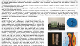
Objectives: This prospective follow-up study aimed to compare the cumulative survival and success rate of implants with bioactive calcium-phosphate coating 7 years after insertion via lateral sinus lift with a transcrestal technique. It also aimed to evaluate bone remodelling around the elevated sinus floor. Methods: Ninety conical self-drilling implants with a calcium phosphate coating (Bioactive) were placed with a simultaneous sinus lift procedure in 40 consecutive male patients between January and December 2008 presenting with 5–6mm of residual sub-sinus bone. Their mean age at the time of surgery was 41 years. Group A (n = 20) were treated with a lateral sinus lifting procedure and simultaneous insertion of 50 implants; the graft was a mixture of platelet-rich fibrin (PRF) and deproteinised bovine bone particles (Bio-Oss) with a collagen membrane (Bio-Gide). In group B, 40 implants were inserted via a transcrestal sinus lift procedure without grafts. The time between implant placement and loading was four months. All restorations were cemented. Primary outcomes were implant survival and success rate, and secondary outcomes were modified Loe and Silness gingival index, Mombelli’s plaque index, and Mühlemann’s bleeding score. The following analyses were used to establish the dates for follow-up: implant primary and secondary stability (using resonance frequency analysis and PeriotestTM) and radiological bone changes around the implants and sinus floor. Results: All patients were seen at 4 and 12 months and followed up at 2–7 years . ISQ dates were collected during implant insertion and after 4 months of healing. The PeriotestTM dates were obtained immediately (4 months, 12 months, 7 years). After healing, ISQ values in group A were 63.1 compared with 59.1 after surgery. In group B, mean ISQ at insertion was 68.8 and 72.1 after 4 months. Mean post-surgical Periotest tm values were –2.15 for group A and −3.64 for group B. After 4 months the value for group A was −4.32 versus −4.74 for group B. The mean Periotest value for both groups after one year was −4.75 and –4.2 after 7 years. Four months after surgery, mean bone loss was –0.52mm in transcrestal cases. After one year, a bone gain of +0.16mm was observed, which decreased to –1. 3mm after 7 years. There was more bone formation after 4 months of surgery (+0.46mm) for the implants in augmented sinuses. After one year, bone loss was –0.4mm; after 7 years it was –2.1mm. The mean gain of bone height in the sinus area in group A was greater (7.8mm) than in group B (3.2mm) after surgery. After rehabilitation, group A had a gain in bone height of 7.6mm, with 7.7mm at 1 year decreasing to 2.3mm at 7 years. For group B the values were 2.9mm at 4 months, 3.1 mm at 1 year, and 3.6mm at 7 years. After 7 years, most cases (75.9%) had normal gingiva. Most implants (93%) had no plaque accumulation, and bleeding could not be provoked in 90%. There were no painful symptoms or mobility in any of the implants. Conclusion: Compared with studies on aftercare for other implant systems, the bioactive calcium–phosphate coated implant system in this study has a promising seven-year survival and success rate. This applies to partially atrophied posterior maxilla edentulous jaws and should be interpreted with respect to the patient selection criteria – transcrestal sinus lift versus lateral approach sinus augmentation with simultaneous implant insertion. The results demonstrate that Bio-Oss is a useful scaffold for augmentation in lateral approach sinus lifts. Transcrestal osteotomy and elevation of the sinus membrane with autogenous local bone is stable, and has an osteoconductive property that allows for direct contact of the implant body with newly formed bone. The resorptive processes in both techniques proceed slowly, providing sufficient time for bone maturation. -
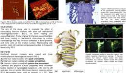
Pectin nanocoating of titanium implant surfaces - an experimental study in rabbits
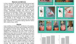
Objectives: The aim of the study was to evaluate the effect of nanocoating titanium implants with plant cell wall-derived rhamnogalacturonan-I, on bone healing and osseointegration. A new strategy to improve osseointegration and bone healing is biochemical stimulation by surface nanocoatings that may increase adhesion of bone proteins, and bone cells at the implant surface. Nanocoating with pectins, plant cell wall-derived polysaccharides, is frequently done using rhamnogalacturonan-I (RG-I). Methods: Machined titanium implants were coated with three modifications of rhamnogalacturonan-I (RG-I). Chemical and physical surface properties were examined before insertion of nanocoated implants (n = 96) into the left and right tibia of rabbits. Machined titanium implants without RG-I nanocoating were used as controls (n = 32). Total number of 128 implants was placed in tibias of 16 rabbits. Fluorochrome bone labels, calcein green and alizarin red S were given intravenously after 9 and 12 days, respectively. The bone response to the nanocoated implants was analyzed qualitatively and quantitatively after 2, 4, 6, and 8 weeks of healing using light microscopy and histomorphometric methods. Results: The RG-I coating influenced the surface chemical composition; wettability and roughness, making the surface more hydrophilic without any major effect on surface micro roughness compared to control implant surfaces.The different modifications of pectin RG-I did not significantly enhance bone healing and osseointegration analyzed after 2, 4, 6, and 8 weeks of healing compared to control implants. Although the qualitative analyses of the fluorochromes indicated a higher activity of bone formation in the mineralization front at the early stage, after 9 and 12 days at the RG-I nanocoated implants compared to the control implants although no significant quantitative difference was demonstrated. Conclusion: The present study showed that nanocoating of titanium implants with pectin RG-Is did not significantly enhance bone healing and osseointegration when placed in rabbit tibia bone. -

RELIGIOUS AND ETHICAL ATTITUDES TO USE OF ALLOGENOUS, XENOGENOUS AND SYNTHETIC TISSUE REPLACEMENT MATERIALS
Objectives: In the field of oral soft and hard tissue reconstruction, there are many data on the use and success of foreign materials of animal, mineral and synthetic origin. Religious and ethical acceptability of these materials is important in our globalised, multicultural world. The aim of our study was to determine the acceptance of allogenic or xenogenic materials among different religious groups and different polytheistics. Methods: Detailed questionnaires were handed out to twelve national leaders of various religious groups: Roman Catholic, Protestant Christian, Muslim, Greek Orthodox, New Apostolic, Romanian Orthodox, Buddhist, Serbian Orthodox, Jewish, Jehova’s Witness and Hindu. The task was to distinguish three groups according[?] to acceptance of different materials by their communities: contraindicated for ethical/religious reasons (group V); informed consent required (group C); and acceptance (group A). At the same time, 120 dentists working in oral surgeries were asked about their awareness of and publicity of ten common materials and their origins, as well as patient-information practices, and patient reactions. Results: All twelve religious groups responded to the questionnaire, mostly after seeking a qualified opinion from an international religious centre. The majority (n = 9) claimed to provide basic information for patients before undergoing any intervention. Some (n = 5) prohibited the use of animal or human tissues for religious or ethical reasons. Others (n = 2) excluded them because of the risk of disease transmission or cruelty to animals. Of the 120 polled dentists or surgeons, the minority (n = 12) were unaware of the origins of the ten described products. For a very small number of patients, the use of tissue replacement materials and the nature of the donor or origin have been important issues, but most did not approve of the use of allogeneic or xenogeneic tissue substitutes in their interventions. Conclusion:Religious attitudes to the preparation and performance of routine dental surgeries showed significant differences. From the findings, it is obvious that patients’ ethical and religious acceptance of biomaterials in this field cannot be assumed. The origin of materials should be clarified and reasonable alternatives pointed out before any intervention to prevent intercultural misunderstanding and judicial consequences. -
Essentials of 3D radiology
Prof. Reinhilde Jacobs and Prof. Marc QuirynenCone beam computer tomography (CBCT) has become an important tool for diagnostics and planning in implant dentistry. This module explains you all the important aspects of the CBCT. -
EVALUATION OF OSTEOGENIC POTENTIAL IN STRO-1-POSITIVE HUMAN TOOTH-GERM DERIVED MESENCHYMAL STEM CELLS (hTGSCs)
Objectives: The objective of this study was compare the osteogenic differentiation potential and mineralisation of STRO-1-positive selected mesenchymal stem cells derived from human tooth germ (hTGSCs) with a heterogeneous hTGSC population, and to determine whether a minor cell subpopulation is more committed to osteogenic differentiation. Methods: Human TGSCs were isolated from dental follicle and apical papilla tissues of impacted tooth germs of third molars from volunteers aged 13–20 years. Cells were expanded and characterised by flow cytometric analysis with CD34, CD45, CD105, CD90, CD44, CD14 and CD117 antigens. Following surface-antigen profiling, STRO-1 antigen, defining a mesenchymal stem cell subpopulation, was used for cell selection with flourescence-activated cell sorting (FACS). Three cell populations (STRO-1 positive, STRO-1 negative, and unsorted) were tested for their ability to differentiate towards osteoblast-like phenotype in osteogenic medium and standard culture medium. Cultures were analysed for cell proliferation (MTS assay and protein assay) and alkaline phosphotase (ALP) activity on days 1, 4, 7, 14 and 21; calcium deposition and gene expression of runx2, osteocalcin and osteonectin with real-time polymerase chain reaction (RT-PCR) on days 7, 14 and 21. Mineralisation and cell phenotype were further screened with Von Kossa staining and confocal microscopy on days 7, 14 and 21. Statistical analysis was made with SPSS Statistics 22[tm]. Kruskal Wallis and Mann–Whitney U tests were applied and a p value of Results: Flow cytometry analysis of passage 3 (p3) hTGSCs showed positivity for CD44, CD90 and CD105, and negativity for CD14, CD45, CD34 and CD117. A total of 11% of p3 hTGSCs were sorted as STRO-1-positive and 24% as STRO-1-negative. In STRO-1-positive cells, osteogenic groups showed significantly higher calcium content than the controls on days 7, 14 and 21. In STRO-1-negative and unsorted cells, the osteogenic groups showed significantly higher calcium content on days 14 and 21 compared to controls. Calcium deposition was not significantly different in osteogenic cell groups in on days 7, 14 and 21. Calcium content seemed to increase in all cell groups from day 7 to day 21. In osteogenic groups, ALP activity of unsorted cells was significantly higher than that of STRO-1-positive and STRO-1-negative groups on days 7, 14 and 21, suggesting a faster rate of osteogenic differentiation in this group, whereas there was no statistical difference between groups on day 21. Activity of ALP was lowest in all groups on day 7 and increased on days 14 and 21. Osteocalcin was expressed in all groups, with no significant difference between them. Osteonectin was highly and significantly expressed in all osteogenic groups on day 7, which is consistent with the findings for ALP and calcium deposition, suggesting that mineralisation was initiated on day 7. Runx2 was downregulated from day 7 to 14. There were no significant differences in the expression of late and early markers of mineralisation in all osteogenic cell groups. Conclusion: Human TGSCs are a novel stem cell source without ethical issues that can be obtained from routine dental treatments. These results suggest they have a high capacityfor osteogenic differentiation. Selecting groups based on STRO-1 negativity or positivity offers no advantages over unsorted cells, but other osteogenic markers might be allow selection of more sensitive osteogenic subpopulations from hTGSCs. -
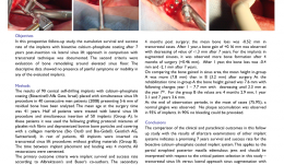
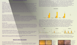
EXPANSION OF THE PERI-IMPLANT ATTACHED GINGIVA WITH A 3-D COLLAGEN MATRIX IN HEAD AND NECK CANCER PATIENTS—RESULTS FROM A PROSPECTIVE CLINICAL AND HISTOLOGICAL STUDY
Objectives: Attached peri-implant gingiva has been proved to influence the integration of dental implants, stable peri-implant soft tissue, and long-term stability of bone leve of osseointegrated dental implants. Methods: The aim of this prospective study was to investigate the 3-D collagen matrix Mucograftä/® for augmentation around dental implants in patients (n = 8) with former head and neck cancer affecting the oral cavity, to enlarge the peri-implant attached gingiva. Clinical performance and shrinking tendency of the matrix were analysed, and biopsies from MG-augmented regions were examined histologically to determine the biomaterial-related tissue-reaction. Results: The participants received a total of 51 dental implants. In all cases, there was uneventful healing without complications or rejection. Six months after vestibuloplasty, there was a mean width of 3.9mm attached peri-implant gingiva with a mean shrinking tendency of 11%. Unrelated to the collagen matrix augmentation, three implants had to be explanted due to peri-implant infection, and dehiscence of the implant windings was observed in two. Histological analysis revealed that the collagen matrix was integrated 8 weeks after vestibuloplasty and augmentation. The compact layer was invaded by mononuclear cells only, towards complete stepwise epithelialisation, and the spongious layer of the matrix was integrated with cell-rich connective tissue involving mononuclear cells alone. Conclusions: Given its limitations, this study revealed that vestibuloplasty with 3-D collagen matrix is a reliable method for enlarging the peri-implant attached gingiva in head and neck cancer patients, with a minimal burden on the patient. Furthermore, the absence of adverse reactions, a favourable tissue reaction, and relatively low shrinking tendency make the collagen matrix a promising alternative to autologous tissue grafts, which are associated with an additional surgical site and post-operative pain. -
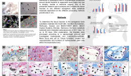
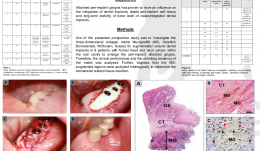
MULTINUCLEATED GIANT CELL FORMATION IN DIFFERENT XENOGENEIC BONE SUBSTITUTE MATERIALS—RESULTS FROM AN IN VIVO STUDY
Objectives: Bone substitute materials have become a reliable alternative to autologous bone in augmentation procedures to regenerate lost bone tissue. From previous investigations it is known that origin, processing and physicochemical material characteristics influence the biomaterial-related tissue reaction. This study aimed to analyse and compare tissue reactions to two different xenogeneic bone substitute materials with different preclinical in vivo processing methods. Methods: To evaluate the tissue reaction to the xenogeneic bone substitute materials Bio-OssTM/® and BEGO-OSSTM/®. The substitute materials were implanted subcutaneously in CD-1 mice for up to 60 days. After explantation, biopsies were processed according to a standardised protocol and investigated histologically and histomorphometrically. The focus was on tissue formation, implant bed vascularisation, and the number of multinucleated giant cells within the implant bed. Results: Both bone substitute materials showed good integration within the peri-implant tissue and no signs of adverse inflammatory effects, and both showed increasing vascularisation over the study period. Within the implant bed of the Bio-Ossä/® substitute, a few multinucleated giant cells were observed on the surface of small-sized bone substitute granules in the early integration period. The tissue reaction to the larger granules in the later stages consisted mainly of mononuclear cells. In contrast, the tissue reaction to the xenogeneic substitute BEGO-OSSä/® consisted of biomaterial surface associated multinucleated giant cells. Histomorphometric analysis confirmed the histological results, with significantly more multinucleated giant cells occurring with BEGO-OSSä/®. The increased numbers of multinucleated giant cells also contributed to a greater vascularisation within the BEGO-OSSä/® implant bed. Conclusions: The two xenogeneic bone substitute materials showed distinct tissue reactions, mainly in the formation and migration of multinucleated giant cells. Both biomaterials have the same origin, thus the observed differences seem to relate to different processing techniques and the resulting physicochemical structures. Induction of multinucleated giant cells is associated with formation of unphysiological amounts of fibrous capsule within the implant bed and this might have a negative outcome on integration of the bone substitute and thus on long-term implant stability. Studies are now needed to investigate the clinical relevance of multinucleated giant cells in human bone tissue regeneration.

Take this survey to help us improve your experience
on Dental Campus
For completion, you get 3 days unlimited access to:
- All lectures
- All cases
- All videos