


-

ACHIEVEMENT OF A HARMONIOUS SMILE VIA HARD AND SOFT TISSUE MANAGEMENTS ACCOMPANYING IMPLANT-SUPPORTED PROSTHESIS. A CASE REPORT.
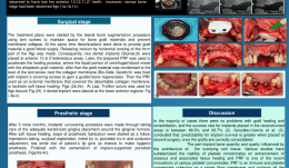
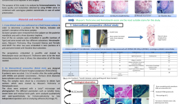
Objectives: A perfect appearance is very important for young patients, especially those who lost their anterior teeth through an accident, because it affects their self-confidence and promotes an active social life. Such patients often need professional treatment and psychological support. Methods: After an accident, a 20-year old women was submitted to the Oral Implantology Department at Istanbul University, seeking a good smile and appearance. Clinical examination revealed the loss of her anterior 13,12,11,21 teeth. Deep bite and narrow bone ridge were also observed. Her treatment plans began with a lateral bone augmentation procedure using platelet-rich fibrin (PRF) as a membrane. Two dental implants were placed at anterior 13 and 21 edentulous areas after 4 months, and after another 3 months, the implants were uncovered with placement of adequate keratinised gingiva around the gingival formers. After soft-tissue healing, prosthesis fabrication was begun and completed by screwing in two screw-retained implant-supported porcelain prostheses. Results: The anterior maxilla was successfully restored by two implant-supported, metal-reinforced porcelain prostheses. Conclusions: Multiple factors must be considered to achieve the good final aesthetic results in the anterior maxilla. The choice of surgical procedure, soft tissue management and prosthetic all affect the outcome in aesthetically challenged trauma patients. -
PLATELET-RICH FIBRIN—A PROMISING DRUG DELIVERY SYSTEM FOR TISSUE REGENERATION IN ORAL AND MAXILLOFACIAL SURGERY: PRECLINICAL AND CLINICAL STUDIES
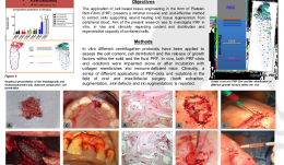
Objectives: The application of cell-based tissue engineering in the form of platelet-rich-fibrin (PRF) is a minimally invasive and cost-effective method to extract cells supporting wound healing and tissue regeneration from peripheral blood. The study aimed to investigate PRF in vitro, in vivo and clinically, in terms of the content, distribution and regenerative capacity of contained cells. Methods: Various in vitro centrifugation protocols have been used to assess cell content and distribution and release of growth factors within solid and fluid PRF. In vivo, both PRF clots and PR solutions were implanted alone or after incubation with collagen membranes into immunodeficient mice. On the clinical side, a range of applications of PRF clots and solutions are reported in tooth extraction, augmentation and decortication in cases of osteonecrosis. Results: The in vitro and in vivo studies clearly showed that changes in centrifuge rotation time and revolutions per minute (r.p.m.) results in different types of cell content. Changing the centrifugation settings also influenced the structure and density of PRF clots. Improved tissue regeneration and healing time were observed in all clinical cases, which were treated with either solid PRF and/or fluid PRF components. Conclusions: PRF can be applied within the operating theatre as a routine-based tissue engineering procedure. Both solid and fluid PRF components can be used clinically for tissue engineering procedures, whereby the surgeon is able to adjust the structure, cell and growth factor content of the PRF constructs to suit a particular clinical need. -
HISTOLOGICAL ANALYSIS OF DIFFERENT COLLAGEN MEMBRANES—PHYSIOLOGICAL OR FOREIGN BODY REACTION?
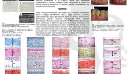
Objectives: Collagen membranes and matrices are known to be reliable barrier materials in guided tissue and guided bone regeneration, for regenerating lost hard and soft tissues in oral and implant surgery. This study presents results from histological investigations of different collagen-based membranes in order to outline the cellular reactions according to specific characteristics of the materials and their manufacturing processes. Methods: Collagen membranes and 3-D collagen matrices that vary in origin and manufacture were implanted subcutaneously in CD-1 mice and Wistar rats, as well as in humans, to determine the tissue reaction within the peri-implant tissue. Samples extracted up to 60 days after implantation were processed and evaluated histologically and histomorphometrically with a special focus on material stability, vascularisation and induction of a multinucleated giant cells triggered foreign-body reactions. Results: Histology revealed distinct differences in the cellular reactions to the various membranes depending on material-specific characteristics and processing techniques. Cellular reactions consisted mainly of mononuclear cells (e.g. macrophages) as sign of high biocompatibility, material integration and lack of a foreign-body reaction, as well as the presence of multinucleated giant cells that arise from macrophages and migrate into the implant bed. Complete volume stability and integrity was also observed in terms of continuous cell and tissue ingrowth and early vascularisation. Conclusions: Collagen-based membranes serve in periodontology and implantology as versatile barriers that separate different tissues and cells during the healing process. For clinical application of collagen membranes, it is very important to have a detailed knowledge of the cell reactions induced in the implant bed. Cellular reactions, volume stability and membrane permeability, as systematically analysed in this study, have a distinct influence on clinical success. -
REHABILITATION OF A PATIENT WITH NOSE MUTILATION RESULTING FROM CARCINOLOGIC SURGERY USING EXTRAORAL FIXTURES AND EPITHESIS
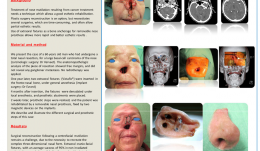
Objectives: Treatment of nose mutilation resulting from cancer treatment requires use of a technique that allows good aesthetic rehabilitation. Plastic surgery is an option, but involves several surgeries, which are time consuming and often have only partial aesthetic results. Using an extraoral fixture for bone anchorage of a removable nose prosthesis results in quicker and better aesthetic results. Methods: We present the case of a 60-year-old man who had undergone a total nasal resection for a large basal cell carcinoma of the nose. Anatomopathologic analysis of the resection showed free margins, and did not reveal any ganglionic metastasis. No radiotherapy was applied. One year later, two extraoral fixtures were inserted in the frontonasal bone, under general anaesthesia, and 4 months after insertion, the fixtures were denudated under local anaesthesia, and prosthetic abutments placed. After 2 weeks, prosthetic steps were realised, and the patient received a removable nasal prosthesis that was fixed by two magnetic devices on the implants. This report describes and illustrates the various surgical and prosthetic steps involved. Results: Patient satisfaction was achieved with this technique. Surgical reconstruction following a centrofacial mutilation is challenging because of the need to recreate a complex 3-D nasal form. Extraoral craniofacial fixtures have an average success rate of 95% in non-irradiated patients and have made it possible for the nasal epithesis to be considered as a forerunner in cosmetic rehabilitation strategies. Conclusions: The clinical case presented here shows that transcutaneous extraoral fixtures may be used as bone anchorage for nasal epitheses, in cases of nasal mutilation resulting from cancer treatment. -

COMPARISON OF PERI-IMPLANT BONE LOSS AT IMPLANTS PLACED INTO ALVEOLAR EXTRACTION SOCKETS FILLED WITH ALLOGRAFT AND PLATELET CONCENTRATES AND AT IMPLANTS PLACED IN NATIVE BONE – A RETROSPECTIVE RADIOGRAPHIC STUDY
Objectives: A loss in height and width of the alveolar process takes place after tooth extraction, and there is consensus on the benefits of post-extractional socket filling. The aim of this study was to compare peri-implant bone loss at implants placed in alveolar sockets filled with particulate allogenous bone graft (DFDBA 300–500µm) and platelet concentrates versus implants placed in native bone. Methods: A retrospective clinical study was performed with 84 patients, in whom 247 implants were placed, 169 of which were in native bone (control group) and 78 in socket-grafted bone (DFDBA 300–500µm) and autogenous platelet concentrates (test group). Peri-implant bone loss was measured by two independent operators at 6 and 12 months after placement. A student’s t-test and ANOVA were used to compare bone loss in test and control groups, and correlation coefficients between the two operators were calculated. Results: The overall mesial and distal peri-implant bone loss was 0.9± 0.7mm and 0.9± 0.8mm, respectively, at 6 months, and 1.0± 0.65mm and 1.1± 0.7 mm at 12 months. In the test group, the bone loss was 0.8 ± 0.8 mm at 6 months and 1.2 ± 0.9 mm at 12 months. In the control group, the bone loss was 1.0 ± 0.75mm at 6 months and 1.02± 0.6 mm at 12 months. There was no statistically significant difference between the two groups or between patients with or without history of periodontitis. However, there was a significant difference between the maxillary and mandibular peri-implant bone loss. Maxillary bone loss was greater than mandibular bone loss by an average of 0.2mm at 6 months and 0.4mm at 12 months. There was also a difference between unitary and total edentations (0.7mm at 6 months, 1.2 mm at 12 months) and partial and total edentations (0.5mm at 6 months, 1.2mm at 12 months). Conclusions: From the results of this study we can conclude that peri-implant bone loss in alveolar sockets filled with allogenous bone graft (DFDBA 300–500µm) and autogenous platelet concentrates are similar to peri-implant bone loss in native bone. -
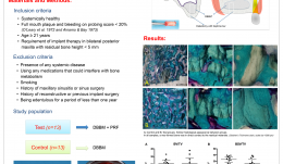
HISTOMORPHOMETRIC EVALUATION OF BONE QUALITY FOR PRESERVATION OF BONE VOLUME AFTER EXTRACTION USING PARTICULATE ALLOGRAFTS (DFDBA) AND PLATELET CONCENTRATES—AN INTERVENTIONAL PROSPECTIVE CLINICAL STUDY
Objectives: There is consensus to preserve bone volume after dental extraction. Some studies show that freeze-dried bone allograft (DFDBA) is the most suitable material for this procedure, and numerous studies support the use of autologous platelet concentrates as an adjuvant in oral surgery. This study aimed to analyse by histomorphometry the quality and maturation of bone obtained using DFDBA alone, or combined with autologous platelet concentrates for socket preservation. Methods: (i) A pre-clinical study on a fresh human cadaver was performed to determine a protocol for fixation, inclusion and staining of bone samples. Six bone samples were removed from the posterior mandibular area with a 4-mm diameter trephine. Four samples were embedded in paraffin (5μm sections) and tested with four different stains: haematoxylin–eosin, Masson’s trichrome, May–Grunewald Giemsa and Periodic Acid Schiff (PAS). Two were embedded in resin (1 μm sections) and tested with toluidine blue. The paraffin-embedded sections stained by haematoxylin–eosin and Masson’s trichrome allowed observation of all the required data. (ii) An interventional prospective clinical study was designed in which eight patients underwent socket grafting with DFDBA and platelet concentrates. After 3–14 months, 2-mm diameter bone cylinders (n = 13) were created with a trephine and sliced in a microtome to obtain 5-μm sections that were stained by Masson’s Trichrome or haematoxylin–eosin. The slices were analysed with a LeicaTM/® microscope and photographed. Evaluated parameters included lamellar bone, woven bone and residual bone graft, and were analysed qualitatively and semiquantitatively by histomorphometry by two independent operators and analysing software (Fiji Image JTM/®). Results: The total new bone area was superior to connective tissue area in all samples (a small proportion of connective tissue and residual DFDBA compared to new bone). Maturation of lamellar bone was observed in 11 out of 12 samples, even after a short post-extraction healing time of 3.5–5 months. Two sections from the same patient also contained bone marrow. Bone remodelling of osteoclasts and osteoblasts occurred in all specimen. No inflammatory infiltrate was detected in any sample. Osteointegration (direct contact with bone) of the DFDBA particles was good in 11 out of 12 samples, and there was progressive resorption and disintegration of the DFDBA particles with time. Fibrocartilage was present in 3 out of 8 patients. Conclusions: Within the limits of these observations on a limited number of patients, it seems that alveolar ridge preservation using particulate DFDBA combined with autologous platelet concentrates allows bone maturation, osseointegration of DFDBA particles, and progressive resorption of DFDBA particles over time. Moreover, fibrocartilage was present in some samples which is an exceptional observation. Further prospective randomised clinical (split mouth) studies are needed with a large number of patients to confirm these observations. -
MAXILLARY SINUS AUGMENTATION WITH PLATELET-RICH FIBRIN (PRF) AND DEPROTEINISED BOVINE BONE MINERAL (DBBM) – A SPLIT-MOUTH HISTOLOGICAL AND HISTOMORPHOMETRIC STUDY
Objectives: To evaluate the effect of platelet-rich fibrin (PRF) in combination with deproteinised bovine bone mineral (DBBM) on bone regeneration in maxillary sinus augmentation. Methods: Thirteen patients (9 men and 4 women aged 49.92 ± 10.37) were enrolled in the study. Twenty-six maxillary sinus augmentation procedures were performed randomly using combined DBBM and PRF (test group) or DBBM alone (control group) in a split-mouth design. In both groups, a resorbable collagen membrane was used to cover the graft material. After 6months, bone biopsies were harvested from the implant sites for histological and histomorphometric evaluation. Clinical and radiographic examinations were performed pre- and post-operatively. Results: There was no qualitative histological difference in the two groups. Neo-formed bone was in direct contact with the residual material in all cases. The amount of newly formed bone (test group 21.38±8.78%; control group 21.25±5.59%), residual bone substitute (test group 25.95±9.54%; control group 32.79±5.89%), and bone substitute in contact with the newly formed bone were similar in both groups (p Conclusions: Both methods were effective for maxillary sinus augmentation. PRF did not improve regenerated bone as well as graft that was incorporated quantitatively. -
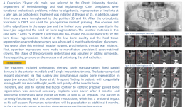
IMPLANT PLACEMENT AND HARD TISSUE REGENERATION FOR OLIGODONTIA AND FOLLOWING ORTHODONTIC THERAPY
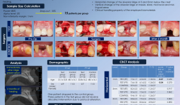
Objectives: Tooth agenesis is one of the most prevalent congenital craniofacial disorders. The term oligodontia is generally used for agenesis of six or more teeth, and it can be related to a syndrome or present as an isolated condition. The prevalence in permanent dentition is 0.14%. This article reports a case of oral rehabilitation using implants and hard tissue regeneration in a patient with agenesis of 15 permanent teeth and following orthodontics. Methods: A 23-year-old Caucasian man, was referred to the Department of Periodontology and Oral Implantology at Ghent University Hospital. His chief complaints were functional and aesthetic, related to oligodontia. Orthodontic treatment was initiated at the age of 15 in preparation for future implant placement. In addition, the upper third molars were transplanted to positions 35 and 45. After orthodontic treatment, a cone beam computerised tomography (CBCT) scan was taken for surgical treatment planning, including implant placement. Concave and knifed-edged bone in the upper jaw and the limited bone quality and quantity of the lower jaw indicated the need for bone augmentation. The materials selected were seven Astra EVä/® implants, and Bio-Ossä/® and Bio-Guideä/® for the hard tissue regeneration. Second-stage surgery for the low bone quality and hard tissue regeneration was scheduled for 6 months after placement. Two weeks after this minimally invasive surgery, prosthodontic therapy was initiated. Open-tray impressions were made to manufacture provisional, screw-retained crowns. The shape of the provisional restorations was adjusted by adding composite, putting pressure on the mucosa and optimising the pink aesthetics. Results: The treatment included orthodontic therapy, tooth transplantation, a fixed partial denture in the anterior mandible and seven single implants. Implant placement was via flap surgery and simultaneous guided bone regeneration (GBR) in the upper jaw. Reduced height, width and quality of alveolar bone are common in patients with congenitally missing teeth are; to deal with this, and to restore the buccal contour, GBR was deemed necessary. Implants were covered after 6 months and provisional restorations were made on teeth and implants. The patients is currently functioning with the provisional restorations, which have had a profound impact on his self-esteem. Permanent restorations will be placed after another 6 months. To date, the buccal contour of the implant sites has shown limited resorption. Conclusions: Treatment of oligodontia patients with dental implants is challenging because of the reduced height, width and quality of alveolar bone. This report demonstrated that a proper multidisciplinary treatment plan offers a predictable solution for complex cases. The choice of biomaterials significantly impacts on the final aesthetic outcome. -
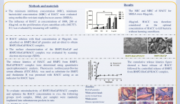
IN VITRO AND IN VIVO CHARACTERISATION OF A NOVEL BONE-DEFECT-FILLING BMP-2-BIOCAP/HACC COMPLEX WITH SEQUENTIALLY ANTIBACTERIAL AND OSTEOINDUCTIVE PROPERTIES
Objectives: Bone grafts with both antibacterial and osteoinductive abilities are greatly needed for treatment of infected bone defects. In this study we developed such a bone graft with a sequential release system: burst release of the powerful antimicrobial agent, hydroxypropyltrimethylammonium chloride chitosan (HACC), followed by sustained release of the osteoinductive agent, BMP-2. Our hypothesis was that this would kill infection-associated bacteria and eventually induce osteogenesis to repair an infected bone defect. Methods: The minimum inhibitory concentration (MIC), minimum bactericidal concentration (MBC) and time–kill curve of HACC against methicillin-resistant Staphylococcus aureus (MRSA) were evaluated. The influence of HACC at concentrations of 1000, 200 or 40μg/mL on the proliferation of pre-osteoblasts was evaluated by measuring cell viability. The influence of 40μg/mL HACC on the BMP-2-induced differentiation of pre-osteoblasts was evaluated by assessing activity of alkaline phosphatase (ALP) , osteocalcin (OCN) expression and mineralisation. BMP-2 was first internally incorporated into biomimetic calcium phosphate granules to form BMP-2-BioCaP. HACC solution was then adsorbed onto its surface to obtain a BMP-2-BioCaP/HACC complex. Confocal laser scanning microscopy (CLSM) was used to observe the structure of BMP-2-BioCaP/HACC using FITC-BSA as a substitute for BMP-2 and staining HACC solution with rhodamine B. The release kinetics of FITC-BSA and rhodamine B were determined by quantitative spectrophotometric analysis. To evaluate osteoinductivity of the complex and to optimise the HACC concentration in vivo, the following groups (n=6) were randomly implanted into subcutaneous pockets in rats: (i) BMP-2-BioCaP/HACC complex (BMP-2-BioCaP/HACC-20, BMP-2-BioCaP/HACC-4 and BMP-2-BioCaP/HACC-0.8 according to content of 20, 4 or 0.8μg HACC); (ii) BMP-2-BioCaP; (iii)BioCaP/HACC; (iv) BioCaP. Five weeks after implantation, samples were retrieved for histological and histomorphometric analysis. Results: The MIC and MBC of HACC for MRSA were 40μg/mL. BioCaP/HACC completely eliminated MRSA within 24 hours. HACC at levels of 1000μg/mL and 200μg/mL (but not 40μg/mL) significantly suppressed cell viability. HACC at a level of 40μg/mL did not significantly influence BMP-2-induced ALP, OCN or mineralisation. Microscopy showed that BMP-2 homogenously distributed throughout BMP-2-BioCaP granules within BMP-2-BioCaP/HACC complex, forming a thin layer of HACC along the surface. The in vitro release profile showed rapid release of HACC, which was completely exhausted after 3 days; BMP-2 was gradually and slowly released, with only about 20% depletion after 30 days. Bone formation was observed only in the BMP-2-containing groups in the in vivo pro-fibrotic environment (subcutaneous sites). In comparison with the positive control group (BMP-2-BioCaP), BMP-2-BioCaP/HACC-4 and BMP-2-BioCaP/HACC-0.8 complexes resulted in similar levels of new bone formation, while BMP-2-BioCaP/HACC-20 was associated with significantly less new bone formation. Conclusions: BMP-2-BioCaP/HACC-4 complex can rapidly eliminate antibiotic-resistant bacteria and efficiently promote new bone formation both in vitro and in vivo – making this a promising material for repairing infected bone defects. HACC is a strong antibiotic that can be readily combined with other bone substitutes, such as Bio-OssTM/® to provide in situ antibacterial activity for bone regeneration associated with infection. -
A RANDOMISED CLINICAL TRIAL FOR RIDGE PRESERVATION USING TWO TYPES OF COLLAGENATED XENOGRAFT/COLLAGEN MEMBRANE—A CONE-BEAM CT STUDY
Objectives: This randomised controlled clinical trial aimed to radiographically compare horizontal and vertical alteration of alveolar ridge following extraction-socket grafting with different combinations of biomaterials: collagenated porcine bone and cross-linked collagen membrane (test group) versus collagenated bovine bone and non-cross-linked collagen membrane (control group). Methods: This study included patients who required at least one anterior or premolar extraction, were aged 20 years old or more, and were able to understand the informed consent. Tooth replacement was planned using either conventional or implant-supported fixed partial dentures. Sample size calculations required 13 patients per group to obtain 80% power at a two-sided alpha level of 0.05%. A total of 30 patients were enrolled, and 15 were randomly allocated to each group. All were blinded to their allocations. Following a single tooth extraction, a collagenated bone substitute was used to fill in the socket, and a collagen membrane was used to cover it. Primary flap closure with several flap designs was attempted. Cone-beam CT (CBCT) scanning was carried out to assess the change in alveolar ridge, once immediately after ridge preservation and once after 4 months of healing. The primary outcome was horizontal alteration of alveolar ridge. Secondary outcomes were vertical alteration of alveolar ridge and clinical handling of the bone substitute and barrier membrane. Results: One patient in the control group dropped out and 3 patients did not receive the allocated intervention in the test group. Intention-to-treat (ITT; control group 14, test group 15) and per protocol (PP; control group 14, test group 12) analyses were performed. In the control and test group, 8 and 7 cases, respectively, showed membrane exposure, secondary healing was uneventful in all cases. There was no statistical difference in ITT and PP analysis between the groups in horizontal alteration of alveolar ridge width, but vertical height in the midfacial area was significantly reduced in the test group. The handling properties of the barrier membrane showed no statistically differences, but the handling properties of the bone substitute was significantly superior in the control group. Conclusions: Ridge preservation using collagenated porcine bone/cross-linked collagen membrane showed similar horizontal ridge alteration to collagenated bovine bone/non-cross-linked collagen membrane. However, vertical ridge changes were more pronounced with the collagenated porcine bone/cross-linked collagen membrane. Further studies including histometric evaluation are required. -
POLYSACCHARIDE NANOGEL CROSS-LINKED MEMBRANE FOR GUIDED BONE REGENERATION (GBR)
Takayuki Miyahara (Japan), Christer Dahlin, Anders Palmquist, Asako Shimoda, Myat Nyan, Yoshihide Hashimoto, Kazunari Akiyoshi, Shohei Kasugai Objectives: GBR is a bone augmentation technique that uses a barrier membrane to create a secluded space for unimpeded bone formation. In a previous report, a wet membrane of polysaccharide nanogel cross-linked (NanoClikä/®) hydrogel was successfully evaluated. The drawbacks of the material are shortshelf-life and difficulties in clinical handling. The purpose of this study was to evaluate the biological outcome of polysaccharide NanoClikä membrane for GBR. Methods: Bilateral symmetrical full-thickness parietal bone defects of 5-mm diameter were created using a bone trephine bur in 16 adult Wistar rats. Each defect was covered with a collagen membrane, a wet or dry NanoClikä membrane, or no membrane (control). The animals were sacrificed at 4 weeks and assessed using micro-CT and histology. Results: New bone formation in terms of bone volume in the bone defect was higher for both membrane groups compared to both control and collagen groups. There was no significant difference between the wet and dry membranes. Notably, the newly formed bone in the defect in both wet and dry groups was uniform and histologically indistinguishable from the original bone, but in the collagen group the new bone showed an irregular structure that was morphologically different from original bone. Conclusions: The dry NanoClik membrane stimulated bone regeneration to the same level as the wet membrane. -
VERTICAL RESORPTION OF AUGMENTED BONE AFTER SINUS LIFTING
Objectives: The use of a bovine-derived xenograft is a reliable alternative to autogenous bone when performing a sinus lift by means of the lateral window technique. Few studies report on the vertical resorption of the augmented bone, and these are mainly based on panoramic images. This retrospective case series aimed to assess vertical resorption of augmented bone by means of peri-apical radiographs. Methods: Between 2012 and 2013, patients underwent sinus lifting in two practices. Each augmentation procedure was performed using the lateral window technique and a bovine-derived xenograft (Bio-Ossä/®). Peri-apical radiographs were taken to assess the following parameters: initial native bone height (BH-B), bone height after sinus augmentation and implant placement (BH-IP), and bone height at tome of evaluation (BH-F). Results: Of a total of 70 eligible patients, 57 (mean age 56 years; SD11) could be evaluated. Mean follow-up time was 19 months (SD9). The parameters BH-B, BH-IP and BH-F were on average 3.87mm (SD1.74), 13.75 mm (SD2.12) and 13.11mm (SD 2.12), respectively. These differences were all highly significant (p Conclusions: Sinus lifting using the lateral window technique and bovine-derived xenografts is a predictable treatment. Irrespective of the initial native bone height, augmentation can be successfully achieved and only limited resorption is likely in the short term.

Take this survey to help us improve your experience
on Dental Campus
For completion, you get 3 days unlimited access to:
- All lectures
- All cases
- All videos