


-
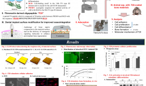

BIOMIMETIC APPROACH TO STIMULATE OSTEOGENESIS ON TITANIUM IMPLANT SURFACES USING FIBRONECTIN-DERIVED OLIGOPEPTIDE
Objectives: Our previous studies demonstrated that a recombinant fibronectin (FN)-derived oligopeptide (F20) stimulated adhesion, proliferation and differentiation of osteoblast in vitro and in vivo. This study aimed to investigate the osteogenic potential of titanium discs coated with F20 for stimulating osseointegration on the surface of dental implants . Methods: The surface characteristics of titanium were assessed using confocal laser scanning microscopy (CLSM). Synthetic F20 was coated onto the titanium discs (machined or SLA) by an adsorption procedure. ST2 cells were then seeded onto the discs, and their adhesion was evaluated using scanning electron microscopy (SEM) and CLSM. Cell proliferation was assessed with PicoGreen staining, and osteoblast differentiation was observed using with real-time polymerase chain reaction (PCR), assessment of alkaline phosphotase (ALP) activity, immunoblot assay and ALP staining. Results: Fluorescence analysis of the fluorescein isothiocyanate (FITC)-labeled F20 disc coating showed good adsorption of F20 and different coating patterns depending on the roughness of the surface. SEM and CLSM showed that the cells were well attached to the machined surface, with greater formation of stress fibres on discs coated with F20 than on uncoated discs. The F20 coating stimulated cellular proliferation, as well as osteoblast differentiation (probably through the extracellular signal-regulated kinase (Erk) signaling pathway). Responses to F20 were slightly better with discs with a machined titanium surface than those with an SLA surface. Conclusion: These results suggest that F20 promotes osteogenesis through the Erk pathway and is a suitable biomolecule for surface modification of dental implants with the purpose of improving osseointegration. -
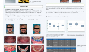
NOBELGUIDE IMPLANT SURGERY AND IMMEDIATE FUNCTION IN FFHB-GRAFTED PATIENTS – A RETROSPECTIVE COHORT STUDY WITH 4–6 YEARS FOLLOW-UP
Objectives: The purpose of this retrospective study was to assess the value of computer-aided technology (NobelGuideä/®) for rehabilitation of fresh-frozen homologous bone (FFHB)-grafted patients with an implant-supported, fixed full-arch prostheses. All patients were treated with a flapless surgical procedure and immediate-function protocol. Implant survival was evaluated after 4–6 years. Methods: Sixty-five patients with atrophic edentulous jaws were reconstructed with FFHB grafts. A NobelGuideä/® flapless surgical protocol was performed for implant placement and all patients received an immediate loaded fixed prosthesis. Implant torque values were recorded at insertion (T1) and after 5 years (T2). Bone levels were assessed radiographically. Soft tissue was examined with a periodontal probe. Pocket probing depth (PPD), modified bleeding index (mBI), keratinised tissue (KT), modified plaque index (mPI) and mucosal plaque scores (MPS) were recorded. Prostheses survival was evaluated. Results: Fourteen men and 51 women were operated on between 2009 and 2011. Of these, 59 (91.7%) were available at a mean follow-up of 62.37 months (range 4–6 years). There were a total of 345 implants (252 Speedy Groovyä/®, 85 MKIIIä/®. Analysis of 321 implants and 72 full-arch prostheses (26 mandibular and 46 maxillary) was performed at follow-up. Survival of implants and prostheses was high at 95.09% and 93.23%, respectively. Most implant and prosthesis failures occurred in the first year. Factors that related significantly to implant failure were smoking, position of the implant at the last distal abutment, and basal maxillary bone fracture. Prosthesis survival was influenced by bruxism and implant failure. Conclusions: This retrospective study demonstrates that computer-guided implant placement in FFHB-grafted patients is a predictable treatment modality that allows clinicians to achieve good primary implant stability, resulting in high survival rates. -

SUPPLEMENTATION OF BIO-GIDE WITH THE SECRETOME OF PLATELETS, ORAL CELLS AND HYPOXIA-MIMETIC AGENTS—EVALUATION OF RELEASE KINETICS AND BIOLOGICAL ACTIVITY IN VITRO
Objectives: Collagen barrier membranes are clinically applied in guided bone regeneration and can be loaded with bioactive factors. We investigated a novel strategy using these membranes as carriers for the secretome of activated platelets (PSEC), oral cells (OSEC) and hypoxia-mimetic agents (HMA). Methods: We measured the release of PSEC, OSEC and HMA from the collagen barrier membrane Bio-Gide. The mitogenic and pro-angiogenic effects were assessed in bioassays with human oral cells. Growth factor release was investigated by immunoassay of the supernatants taken from Bio-Gide. The release of HMA was assessed by photometry. Results: Bio-Gide releases PSEC, OSEC and HMA with a burst-like release kinetic. The release of total protein and growth factors such as platelet-derived growth factor (PDGF-BB(, transforming growth factor(TGF)-β, and vascular endothelial growth factor (VEGF) peaked in the first hours and rapidly decreased thereafter. A similar release kinetic was observed with HMA. Supernatants of PSEC-loaded membranes stimulated proliferation. The effect of PSEC-loaded Bio-Gide on proliferation decreased rapidly, as observed in supernatants taken after 3, 6, 24 and 48 hours. Supernatants from Bio-Gide loaded with secretome from unwashed platelets had a stronger impact on proliferation than that from washed platelets. Bio-Gide loaded with HMA induced a strong pro-angiogenic response, which was lower with supernatants taken after 3, 6, 24 and 48 hours. Conclusion: Bio-Gide releases PSEC, OSEC and HMA with a burst-like release kinetic. The ability of PSEC- or HMA-loaded collagen barrier membranes stimulate oral tissue regeneration through their mitogenic and pro-angiogenic effects should be assessed in preclinical studies. -
USE OF 3-D COLLAGEN MATRIX (MUCOGRAFT) IN SURGICAL MANAGEMENT OF ORO-ANTRAL COMMUNICATION (OAC) AND ITS COMPARISON TO OTHER MODALITIES
Objectives: Oro-antral communications (OAC) can occur after extraction of teeth in the maxilla. Several closure techniques can be used to treat the complication, including conservative flapless techniques. The aim of this study was to compare different techniques for closure of OAC and evaluate their efficiencies. Methods: Ninety-five patients with OAC were divided into five groups of 15 patients for treatment with: a flapless technique with Mucograft Sealä/®, a flapless technique with collagen in root design, a Wasmundä/® buccal flap, free-flap from palate, and a sandwich technique (collagen–Bio-Ossä/®-collagen-free gingival graft). In the first 2 weeks after intervention, patients recorded their perceptions of bleeding, swelling, pain and bruising on a visual analogue scale (VAS). Post-surgical complications were assessed clinically at 1 week, 2 weeks, 1 month and 6 months post-surgery. Results: Closure of all wounds was achieved from 2 weeks to 1 month. There were no post-surgical complications. All VAS parameters decreased to almost zero by day 14 and were maintained after 6 months. Scores for swelling and bruising peaked on days 2 and 3, respectively, before decreasing. Conclusions: Using a flap or flapless technique depends on personal choice and skills of the surgeon. The use of Mucograftä/® for closure of OAC is effective and safe, and is associated with low morbidity and no post-surgical complications. -
REGENERATION POTENTIAL OF BLOOD MESENCHYMAL STEM CELLS – A SIMPLE PROTOCOL FOR BONE GRAFTS IN MEDICAL OR DENTAL OFFICES AND SOFT TISSUE OR CARTILAGE REGENERATION
Objectives: Mesenchymal stem cells are considered to have a positive impact on tissue regeneration, but they require many manipulations and invasive procedures, such as cell harvesting from bone marrow. This study aimed to evaluate a protocol for using smart blood concentrate in the form of injectable platelet rich fibrin (i-PRF) provides platelets, inflammatory cells and considerable quantities of mesenchymal stem cells from a simple blood withdrawal procedure with a short spin time. Methods: Autologous blood was withdrawn and centrifuged in specific i-PRF tubes at a very low speed for a short time, producing a supernatant of i-PRF. After flow cytometric analysis, specific markers (CD34–, CD45–, CD44+, CD73+, CD90+, CD105+) were applied to detect mesenchymal stem cells. The supernatant was injected into human knee and temporomandibular (TMJ) joints and in sites for soft tissue and bone regeneration within the oral cavity in a total of 40 patients suffering from TMJ and knee joint disorders. Results: Numerous mesenchymal stem cells (0.4–2.0% of total cells) were present in the supernatant. They were cultured in in mono-culture and co-culture with other mesenchymal cells, such as osteoblasts, fibroblasts and endothelial cells. Injection into a graft included clotting of granulesin 1 minute and produced a solid bone graft without any granule mobility (sticky bone graft). Six injections resulted in cartilage regeneration in 10 patients and significant pain relief was achieved in 10 with TMJ dysfunction. In the other 20, better soft tissue and bone regeneration was achieved compared with control groups who received bone substitute and collagen-based materials without i-PRF. Conclusions: This simple protocol may lead to new clinical applications of stem cells for tissue regeneration. The presence of stem cells within the inflammatory milieu might optimise synergy between mesenchymal stem cells and inflammatory cells for soft tissue and bone regeneration. -
EXTRACTED TOOTH ROOTS USED FOR LATERAL ALVEOLAR RIDGE AUGMENTATION—A PROOF-OF-CONCEPT STUDY
Objectives: To assess the efficacy of tooth roots used as autografts for lateral ridge augmentation and two-stage early osseointegration of titanium implants. Methods: The maxillary premolars of foxhounds (n=8) were randomly assigned to undergo either endondontic therapy (PM-E) or be untreated (PM-C). Retromolar cortical autogenous bone (AB) blocks served as controls. PM-E, PM-C and AB were used for ridge augmentation at chronic-type defects in both lower quadrants. After 12 weeks, titanium implants were inserted and left to heal for another 3 weeks. Histological analyses focused on the crestal ridge width (CW), augmented area (AA) and bone-to-implant contact (BIC). Results: Both PM and AB grafts with exposures 3 (AB), 4 (PM-C) and 7 (PM-E) were gradually involved in the bone remodelling process and were associated with replacement resorption. Median crestal ridge widths were 2.70mm (PM-C) vs 3.30 mm (AB) and 2.96mm (PM-E) vs 3.35 mm (AB). For augmented areas they were 7.55mm2 (PM-C) vs 8.56 mm2 (AB) and 11.20mm2 (PM-E) vs 6.60 mm2 (AB). For bone-to-implant contact they were 36.96% (PM-C) vs 64.10% (AB) and 50.79% (PM-E) vs 32.53% (AB). These values were comparable in both PM and AB groups (p>0.05). Conclusions: The extracted tooth roots have structural and biological potential as alternative autografts to autogenous bone. A higher exposure rate may be expected when using endodontically treated teeth. -
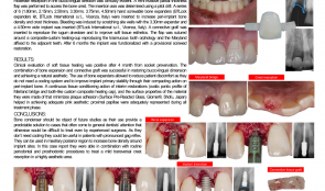
BONE EXPANSION WITH SCREWABLE BONE EXPANDERS IN THE AESTHETIC ZONE – A CASE REPORT
Objectives: Ridge-splitting techniques are used in resorbed ridges. They are difficult to perform and highly technique sensitive. In this case report we present a miniinvasive protocol for a safe and reproducible single-implant rehabilitation in resorbed anterior sites. The rationale for our technique is increasing crest thickness with a controlled bone expansion and achieving optimal aesthetic with a safe and simple protocol. Methods: An 18-year-old patient had a traumatic avulsion of the right central incisor due to maxillofacial trauma. Surgical debridement of the contaminated alveolus and socket preservation using deproteinised bovine bone were performed. An epithelial connective graft was sutured to size to cover the bone graft and promote healing by first intention. A Maryland bridge was used for interim restoration during the healing period, using a woven polyethylene fibre, the lost tooth and a flowable composite to define an ovatic pontic profile (BeautifilTM/®). After 4 months the site was healed. The proximal papillae were maintained and moderate resorption in the buccolingual direction was clinically evident. A mini-invasive partial thickness flap was performed to access the bone crest, with an insertion axis determined using a pilot drill. A series of four ( 1.80mm, 2.15mm, 2.50mm, 3.30mm) hand-screwable bone expanders (BTLockTM/® expanders kit) were inserted to increase peri-implant bone density and crest thickness. Bleeding was induced by scratching site walls with the 2.50-mm expander, and a 3.75-mm wide implant was inserted. A connective graft was inserted to reproduce the iugum alveolare and to improve soft tissue aesthetics. The flap was sutured around a composite custom healing cup, reproducing the transmucous tooth morphology and the Maryland was affixed to the adjacent teeth. After 6 months the implant was functionalised with a provisional screwed restoration. Results: Clinical evaluation of soft tissue healing was positive after 4 months from socket preservation. The combination of bone expansion and connective graft was successful in restoring the buccolingual dimension and achieving a natural aesthetic. The use of bone expanders reduced patient discomfort because no cooling system is needed, and improved implant primary stability through their compacting action on peri-implant bone. An adequate pink aesthetic was achieved by using a continuous tissue-conditioning action of interim restorations with an ovatic pontic profile of the Mariland bridge and a tooth-like custom composite healing cap, and surfaces with properties that minimise plaque adhesion (Surface Pre-Reacted GlassTM/®). Proximal papillae were adequately represented during all treatment phases. Conclusions: Bone condensers should be the object of future studies as their use provides a predictable solution for cases that often come to the attention of general dentists but are difficult to treat (even for experienced surgeons). Because they do not need cooling, they are useful in patients with a pronounced gag reflex. They can be used in the posterior maxillary region to increase bone density around implant sites. In this report, they were used in combination with routine periodontal and prosthodontic procedures to treat a mild transversal crest resorption in a highly aesthetic area. -
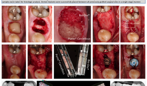
CLINICAL AND HISTOLOGICAL ANALYSIS OF DUAL SOCKET GRAFTING – A PILOT STUDY
Objectives: Extraction socket grafting has been validated for preservation of alveolar ridge dimensions following tooth loss. For making a scientific comparison of different materials requires extraction of at least two teeth. This pilot study aimed to explore the viability of using a single mandibular molar extraction to compare two different materials. The results were evaluated and compared clinically, radiographically and histologically. Methods: Five consecutive patients received an atraumatic tooth extraction of the mandibular first molar with subsequent placement of bone allograft (PurosTM/®) in one socket and xenograft (Bio-OssTM/®) in the other. All sites were covered with a single layer of an absorbable collagen membrane (Ossix PlusTM/®), which was intentionally left exposed. After 20 weeks, the surgical sites were accessed for placement of dental implants. Prior to placement, two trephine core samples were taken for histologic analysis. Dental implants were successfully placed between all previously grafted surgical sites in a single-stage manner. Results: Complete healing of both grafted sites was observed at 20 weeks. The clinical and radiographic aspects of the sites grafted with either allograft or xenograft were significantly different. Xenograft-grafted sites were significantly more radiopaque and residual graft particles were seen at the alveolar crest; allograft-grafted sites demonstrated radiopacity similar to non-grafted adjacent areas, and no visible residual graft particles were seen at the osseous crest, suggesting a higher degree of material turnover. Significant differences were also observed histologically, with more residual graft particles and less vital bone where xenograft was placed, confirming that the two materials behave in significantly different ways when placed in extraction sites for alveolar ridge preservation. Conclusions: This pilot study demonstrated that a single mandibular molar socket can be used to make both clinical and histological comparisons between two different types of bone grafting materials. Future studies should consider using this less traumatic and more convenient research protocol. -
![IN VITRO ASSESSMENT OF PRIMARY STABILITY OF BONE TRUST[TM] SINUS IMPLANT DESIGN—A PRELIMINARY STUDY](https://library.dental-campus.com/media/catalog/product/cache/3/small_image/295x174/17f82f742ffe127f42dca9de82fb58b1/s/i/sinus_implantat.pdf_1_2.png)
IN VITRO ASSESSMENT OF PRIMARY STABILITY OF BONE TRUST[TM] SINUS IMPLANT DESIGN—A PRELIMINARY STUDY
Objectives: The aim of this in vitro study was to analyse the primary stability of BoneTrust Sinus[tm] implant, which suggested to enable higher primary stability by its special design with reduced thread section in cases of reduced vertical bone height, in comparison with standart BoneTrust[tm] implants. Methods: A bone window with dimensions of 3cm x 4cm x 3cm, resembling the maxillary bone window of the lateral sinus wall, with a 4-mm residual bone height was prepared at the dorsal side of freshly slaughtered bovine ribs. Each bone window was fitted with either a BoneTrust Sinus[tm] implant (Sinus) or a Standard BoneTrust[tm] implant (Standard) with the same diameter (either 4mm or 5mm). After implant placement, the international stability quotient (ISQ) was measured by using radiofrequency analysis (RFA) with the Osstell device. Results: A total of 88 implants were placed. ISQ values were 71–84 (Sinus) and 64–80 (Standard) with diameters of 4mm, and 63–78 (Sinus) and 64–80 (Standard) with diameters of 5mm. Among the implants with 4-mm diameters, all Sinus implants showed higher ISQ values than Standard implants (p Conclusion: Within the limitations of this in vitro study using bovine ribs as an experimental model, higher implant primary stability was found for the BoneTrust Sinus[tm] implants with a 4-mm diameter compared to Standart BoneTrust[tm] implants. The use of BoneTrust Sinus[tm] implants with a diameter of 4mm may be associated with higher ISQ values during simultaneous implant placement in conjunction with lateral sinus floor augmentation as suggested by the manufacturer. -
PROTEASE-ACTIVATED RECEPTOR-1 AND PERIODONTAL TISSUE REPAIR
Objectives: Protease-activated receptor-1 (PAR-1) activation by thrombin may play a role in bone growth, repair and homeostasis of periodontal tissues. The aim of this study was to investigate PAR-1 expression in patients with periodontitis, before and after non-surgical periodontal treatment, and relate its expression with the presence of inflammatory biomarkers. Methods: Gingival crevicular fluid (GCF) samples were collected and clinical evaluations made (probing depth, clinical attachment level, bleeding on probing, gingival and plaque indices) in periodontally healthy subjects and patients with chronic periodontitis (CP) at baseline and 6 weeks after periodontal treatment. PAR-1 mRNA expression at the GCF was evaluated by qPCR. Flow cytometry analysis identified the PAR-1-expressing cells. GCF inflammatory biomarkers were also assessed. Results: After periodontal treatment, clinical parameters were significantly improved (p Conclusions: Periodontal treatment resulted in PAR-1 overexpression in the GCF, and PAR-1 expression was inversely associated with the expression of inflammatory biomarkers in the GCF. Thus, the data suggest the importance of PAR-1 mediating the known anabolic actions of thrombin in the periodontium. More studies are necessary to elucidate the mechanism of PAR-1 on periodontal tissue repair.

Take this survey to help us improve your experience
on Dental Campus
For completion, you get 3 days unlimited access to:
- All lectures
- All cases
- All videos