


-
- English
- Deutsch
- Spanish
-
- General Diagnostics
- Treatment
- Tools/Materials
- Basic/General Knowledge
- Research
- Events
- Organisations
-
-
-
-
-
-

-

-
Innovative CAD/CAM treatment approaches for implant-supported fixed restorations
Beuer, Florian / Stimmelmayr, Michael / Schweiger, JosefOutline: - Patient presentation - Preparing the implant bed, implant placement, checking implant positions - Securing the insertion posts to the index for fabrication of the cast - Suturing details - Delivery of the adapted long-term provisional - Fabricating the cast and the gingival mask, transferring the pontic emergence profiles to the gingival mask, mask adaptation - The master cast under the strip scanner with scan bodies on the laboratory analogs - CAD crown design and virtual anatomic shaping - CAM fabrication of a zirconia abutment - Adhesively connecting the zirconia abutment to the titanium base - Reentry, split-thickness flap, vestibuloplasty, connecting the zirconia abutments to the implants - Mucosal graft to restore a soft-tissue defect - Intraoral impression of the abutments - Fabricating the definitive lithium disilicate crowns: virtual crown design; CAM milling, characterization of the crowns - Delivery, final adjustments, presentation of the treatment outcome -
Rational implantation (individual implant in 20 min.)
Bücking, Wolfram -
-
Implant placement in the lower posterior area with immediate provisionalization
Hürzeler, Markus B. -
SOS - An innovative method for the implantological rehabilitation of the edentulous mandible
Sliwowski, Christoph T. -
-
Surgical Treatment of Periodontitis Using a Minimally Invasive Approach
Beck, FrankThis case is an excellent demonstration of the use of the minimally invasive access flap technique for treatment of (chronic) periodontitis in an esthetically critical zone. The access flap was used in conjunction with enamel matrix proteins for regenerative therapy., -
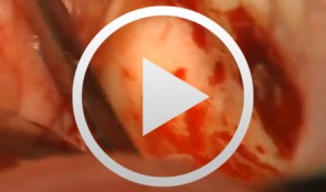
Microsurgical Removal of a Foreign Body from the Mandibular Canal
Schultze-Mosgau, StefanOverview: - Access and incision: Creation of a vestibular pedicled mucoperiosteal flap via a gingival margin incision while preserving the papilla - Removal of vestibular bone in the region of tooth 46 using a microsurgical instrument - Exposure of the neurovascular bundle - Removal of the foreign body - Re-adaptation of the mucoperiosteal flap - Wound closure with atraumatic suture material Contents: Female patient with an indication for microsurgical foreign body removal (removal of a fractured root canal instrument from a previous endodontic treatment of tooth 46) using a surgical microscope. The foreign body extends from the apex into the mandibular canal. -
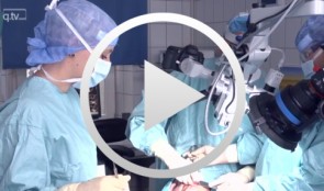
Microsurgical lateral sinus floor elevation (LSFE)
Nölken, RobertOutline: - Incision - Flap mobilization - Lateral sinus fenestration - Elevation of the Schneiderian membrane - Implant bed preparation - Bone chip harvesting at the mandibular angle - Filling of sinus lift lumen with autologous bone chips - Implant insertion - Covering the lateral sinus cavity with collagen membrane - Wound closure List of materials - Zeiss Pro Dent microscope with beam splitter and Panasonic 3 CCD camera - Scalpel holder (Ustomed) with Swann-Morton blades 15C and 12D - Narrow rasp (Hu-Friedy) - Micro-vacuum (Luer Lock Suction Tip, American Dental Systems) - Disposable vacuum tube set (Bexamed) - Disposable draping, Lindau (Aescologic) - Piezosurgery with diamond ball (Mectron) - Microforceps (Hu-Friedy) - Excavator (Martin) - Periodontometer, 1-mm gradation (Hu-Friedy) - OsseoSpeed implant set, Dentsply Implants: Marking drill; Twist drill, 2 mm; Depth gauge; Pilot drill, 2/3.2 mm; Twist drill, 3.2 mm; Tapered drill, 3.2/5 mm; OsseoSpeed TX implant, 5.0 × 11 mm; Closure screw, 4.5/5 mm - Columbia curette (Ustomed) - Micross scraper (Meta) - Needle holder (Ustomed) - Langenbeck wound retractor (Ustomed) - Kelly scissors (Ustomed) - Buchanan endodontic hand plugger (American Dental Systems) - Resorbable collagen membrane (Resodont, Resorba) - Ethilon 5-0 FS-3 (Ethicon) - Prolene 6-0 DA-2 (Ethicon)